. 2014 Oct;27(3-4):163-74.
doi: 10.1293/tox.2014-0007. Epub 2014 May 26.

Green Tea Extract-induced Acute Hepatotoxicity in Rats

Affiliations

Green Tea Extract-induced Acute Hepatotoxicity in Rats

Yuko Emoto et al. J Toxicol Pathol. 2014 Oct.

Erratum in

  • Errata (Printer's correction).
    [No authors listed] [No authors listed] J Toxicol Pathol. 2016 Jan;29(1):74. Epub 2016 Feb 17. J Toxicol Pathol. 2016. PMID: 26989306 Free PMC article.

Abstract

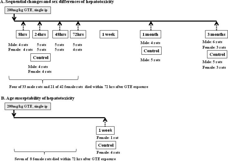
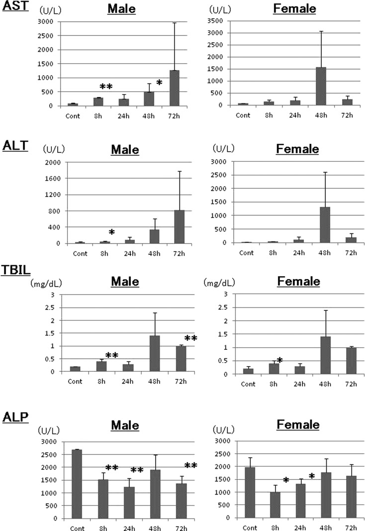
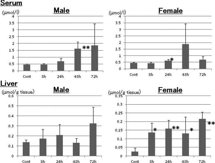
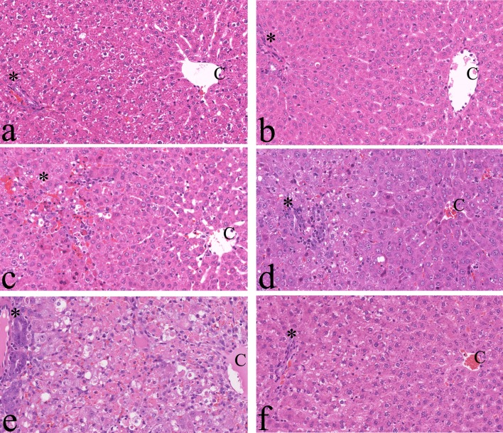
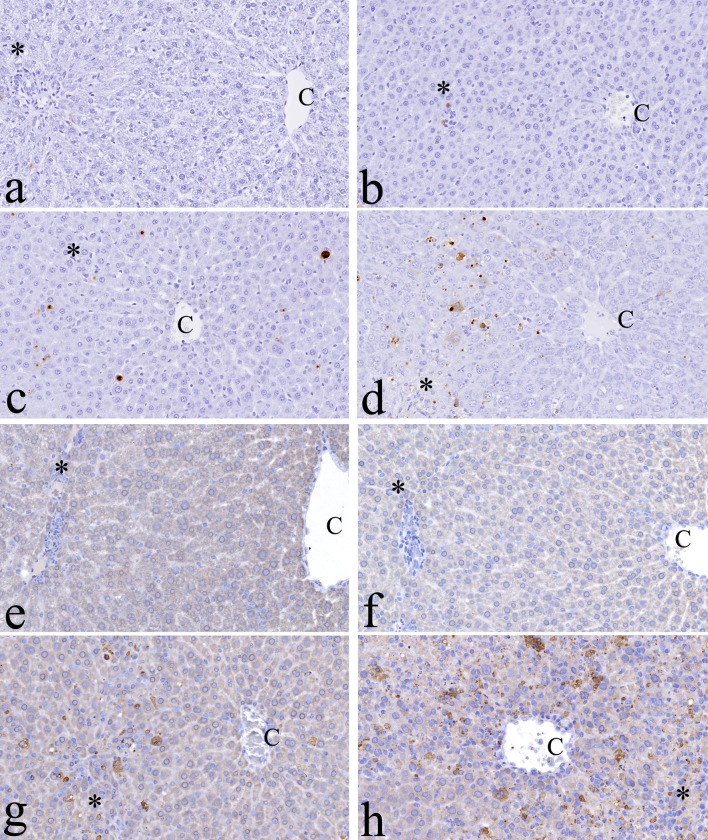
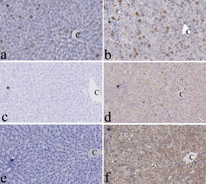
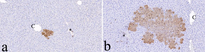
Although green tea is considered to be a healthy beverage, hepatotoxicity associated with the consumption of green tea extract has been reported. In the present study, we characterized the hepatotoxicity of green tea extract in rats and explored the responsible mechanism. Six-week-old IGS rats received a single intraperitoneal (ip) injection of 200 mg/kg green tea extract (THEA-FLAN 90S). At 8, 24, 48 and 72 hrs and 1 and 3 months after exposure, liver damage was assessed by using blood-chemistry, histopathology, and immunohistochemistry to detect cell death (TUNEL and caspase-3) and proliferative activity (PCNA). Analyses of malondialdehyde (MDA) in serum and the liver and of MDA and thymidine glycol (TG) by immunohistochemistry, as oxidative stress markers, were performed. Placental glutathione S-transferase (GST-P), which is a marker of hepatocarcinogenesis, was also immunohistochemically stained. To examine toxicity at older ages, 200 mg/kg green tea extract was administered to 18-wk-old female rats. In 6-wk-old rats, 12% of males and 50% of females died within 72 hrs. In 18-wk-old rats, 88% died within 72 hrs. The serum levels of aspartate aminotransferase, alanine aminotransferase and/or total bilirubin increased in both males and females. Single-cell necrosis with positive signs of TUNEL and caspase-3 was seen in perilobular hepatocytes from 8 hrs onward in all lobular areas. PCNA-positive hepatocytes increased at 48 hrs. MDA levels in the serum and liver tended to increase, and MDA- and TG-positive hepatocytes were seen immunohistochemically. GST-P-positive hepatocellular altered foci were detected in one female rat at the 3-month time point. In conclusion, a single injection of green tea extract induced acute and severe hepatotoxicity, which might be associated with lipid peroxidation and DNA oxidative stress in hepatocytes.

Keywords: apoptosis; green tea; hepatotoxicity; oxidative stress; rats.


Figures

Fig. 1.
Fig. 1.
Experimental protocol. Solid arrows, injection of green tea extract (GTE); broken arrows, time points of necropsy.
Fig. 2.
Fig. 2.
Blood chemistry. The serum concentrations of aspartate aminotransferase (AST), alanine aminotransferase (ALT), alkaline phosphatase (ALP) and total bilirubin (TBIL) were analyzed. *: p<0.05; **: p<0.01.
Fig. 3.
Fig. 3.
Analysis of malondialdehyde in serum and the liver. *: p<0.05; **: p<0.01.
Fig. 4.
Fig. 4.
Histopathological findings. a, Control; b–f, 8 hrs, 24 hrs, 48 hrs, 72 hrs and 1 month after green tea extract (GTE) exposure, respectively. Note that hepatocellular damage progresses gradually from the perilobular (c) to centrilobular area after GTE exposure (e). C: central vein. *Glisson’s sheath. H & E, ×200.
Fig. 5.
Fig. 5.
Immunohistochemistry for cell death in the liver. The signals of terminal deoxynucleotidyl transferase-mediated d UTP digoxigenin nick and end-labeling (TUNEL) can be in the nuclei of hepatocytes of green tea extract (GTE)-exposed rats. a, Control; b–d, 8 hrs, 24 hrs and 48 hrs after GTE exposures, respectively. Caspase 3 signals can also be seen in the nuclei of hepatocytes of GTE-exposed rats. e, Control; f–h, 8 hrs, 24 hrs and 48 hrs after GTE exposure, respectively. C: central vein. *Glisson’s sheath. ×200.
Fig. 6.
Fig. 6.
Immunohistochemistry for proliferative activity and oxidative stress in the liver. Proliferating cell nuclear antigen (PCNA) signals can be seen in the nuclei of hepatocytes of control (a) and green tea extract (GTE)-exposed rats (b). Note that more positive cells can be seen in the rats 48 hrs after GTE exposure (b). No thymidine glycol (TG) signals can be seen in control rats (c), while TG signals in the nuclei of hepatocytes were detected in the rats 48 hrs after GTE exposure (d). No malondialdehyde (MDA) signals can be seen in control rats (e), while positivity for MDA in the hepatocellular cytoplasm was detected in the rats 48 hrs after GTE exposure (f). C: central vein. *Glisson’s sheath. ×200.
Fig. 7.
Fig. 7.
Immunohistochemistry for placental glutathione S-transferase (GST-P). Note that the larger GST-P-positive foci are found 3 months after green tea extract (GTE) exposure (b) compared with the age-matched controls (a). Bile duct epithelial cells are normally positive for GST-P in both groups. C: central vein. *Glisson’s sheath. ×100.

References

    1. Yuan JM, Sun C, and Butler LM. Tea and cancer prevention: epidemiological studies. Pharmacol Res. 64: 123–135. 2011. - PMC - PubMed
    1. Chow HHS, and Hakim IA. Pharmacokinetic and chemoprevention studies on tea in humans. Pharmacol Res. 64: 105–112. 2011. - PMC - PubMed
    1. Lambert JD, and Elias RJ. The antioxidant and pro-oxidant activities of green tea polyphenols: a role in cancer prevention. Arch Biochem Biophys. 501: 65–72. 2010. - PMC - PubMed
    1. Cooper R, Morré DJ, and Morré DM. Medicinal benefits of green tea: part II. review of anticancer properties. J Altern Complement Med. 11: 639–652. 2005. - PubMed
    1. Cooper R, Morré DJ, and Morré DM. Medicinal benefits of green tea: Part I. Review of noncancer health benefits. J Altern Complement Med. 11: 521–528. 2005. - PubMed