Review
. 2014 Nov;29(6):685-702.
doi: 10.3904/kjim.2014.29.6.685. Epub 2014 Oct 31.

Role of modern 3D echocardiography in valvular heart disease

Affiliations
Review

Role of modern 3D echocardiography in valvular heart disease

Takahiro Shiota. Korean J Intern Med. 2014 Nov.

Abstract

Three-dimensional (3D) echocardiography has been conceived as one of the most promising methods for the diagnosis of valvular heart disease, and recently has become an integral clinical tool thanks to the development of high quality real-time transesophageal echocardiography (TEE). In particular, for mitral valve diseases, this new approach has proven to be the most unique, powerful, and convincing method for understanding the complicated anatomy of the mitral valve and its dynamism. The method has been useful for surgical management, including robotic mitral valve repair. Moreover, this method has become indispensable for nonsurgical mitral procedures such as edge to edge mitral repair and transcatheter closure of paravaluvular leaks. In addition, color Doppler 3D echo has been valuable to identify the location of the regurgitant orifice and the severity of the mitral regurgitation. For aortic and tricuspid valve diseases, this method may not be quite as valuable as for the mitral valve. However, the necessity of 3D echo is recognized for certain situations even for these valves, such as for evaluating the aortic annulus for transcatheter aortic valve implantation. It is now clear that this method, especially with the continued development of real-time 3D TEE technology, will enhance the diagnosis and management of patients with these valvular heart diseases.

Keywords: Aortic valve; Echocardigraphy; Mitral valve; Three-dimensional.


Conflict of interest statement

No potential conflict of interest relevant to this article was reported.

Figures

Figure 1
Figure 1
Normal mitral valve imaged by 2-dimensional (D) echo (A, a long axis view in diastole; B, in systole) and real-time 3D transesophageal echocardiography (C, a view from the left atrium in systole; D, in eerily diastole; E, in late diastole). Arrows indicate each leaflet: lateral P1, middle P2, and medial P3. Ao, aorta; LA, left atrium; LV, left ventricle.
Figure 2
Figure 2
Mitral valve prolapse imaged by (A) 2-dimensional (D) transthoracic echocardiography (arrow) and (B) real-time 3D transesophageal echocardiography (arrows). LA, left atrium; LV, left ventricle; RA, right atrium; RV, right ventricle.
Figure 3
Figure 3
Two types (A, one continuous jet; B, two separate jets) of functional mitral regurgitation jet clearly distinguished by color Doppler 3-dimensional transesophageal echocardiography.
Figure 4
Figure 4
Difference in the shape of vena contracta from degenerative mitral valve disease (Ac) and functional mitral valve disease (Bc) delineated by color Doppler 3-dimensional transesophageal echocardiography.
Figure 5
Figure 5
Mitral valve and residual small regurgitant jet (an arrow) imaged by 3-dimensional transesophageal echocardiography (A) without and (B) with color Doppler after clip procedure. LV, left ventricle.
Figure 6
Figure 6
A residual mitral regurgitation immediately after surgical mitral valve replacement (a tissue valve). (A) The upper panel shows 2-dimensional (D) transesophageal echocardiography (TEE) image. (B) Color Doppler 3D TEE image could demonstrate the exact location of the residual mitral regurgitation (MR) (arrow) which could assist in second pump correction of the MR. AP, appendage; AV, aortic valve; MV, mitral valve.
Figure 7
Figure 7
Three-dimensional transesophageal echocardiography images of a stenotic mitral valve (A) from the left atrium and (B) from the left ventricle. Arrows indicate severe calcifications.
Figure 8
Figure 8
Three-dimensional transesophageal echocardiography images of normal aortic valve in a cardiac cycle (A, diastole; B, early systole; C, mid-systole).
Figure 9
Figure 9
Three-dimensional transesophageal echocardiography images of an aortic valve with severe aortic valve regurgitation. (A) Panel shows the valve in mid systle, (B) panel in end systole, and (C) panel in diastole. Arrows indicate a large regurgitant orifice viewed from the ascending aorta.
Figure 10
Figure 10
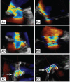
The measurement of aortic valve area by three-dimensional transesophageal echocardiography. (A, B) The tip of the aortic valve was obtained as the smallest possible area. (C, D) The shape and area of the aortic valve changed in a trivial different plane from the tip. The dotted lines indicate aortic valve area at each level. Ao, aorta; LA, left atrium; LV, left ventricle; RA, right atrium.
Figure 11
Figure 11
Real-time three-dimensional transesophageal images, showing the difference in the shape of left ventricular outflow tract (LVOT) between discrete subaortic stenosis (DSS) and hypertrophic obstructive cardiomyopathy. (A) The DSS images show the almost oval or flat shape of the LVOT (Ad) and subaortic membrane with small fenestration at left upper site of membrane (Aa and Ab, arrow). The thin membranous structure changes its angle to decrease the LVOT area along the blood stream (Ac and Ad, arrows). (B) In hypertrophic obstructive cardiomyopathy, the shape of the LVOT is a V formation or two separate open spaces due to systolic anterior motion of mitral anterior leaflet (Bc and Bd, arrows).
Figure 12
Figure 12
Three-dimensional transesophageal close up views of the normal tricuspid valve in a cardiac cycle. (A) Panel shows the tricuspid valve in end-diastole, (B) in mid-diastole, (C) in early diastole, and (D) in systole. A, anterior cusp; P, posterior cusp; S, septal cusp.
Figure 13
Figure 13
Reconstructed normal tricuspid annulus with the use of (A) 3-dimensional (D) echocardiography and (B) its application to the development of a new 3D ring for surgical annuloplasty. A, anterior; L, lateral; P, posterior; S, septal.
Figure 14
Figure 14
(A) Upper panel shows 2-dimensional (D) transesophageal (TEE) images without (left) and with (right) color Doppler in a patient with thrombus formation on the mechanical mitral valve (arrow). The lateral leaflet does not open. Lower panels show real-time 3D TEE images from the same patient, (B) left in systole and (C) right in diastole. A large thrombus is well visualized with 3D TEE (arrows).
Figure 15
Figure 15
(A) En-face view of mitral valve with a vegetation attached to both anterior and posterior leaflets. Arrow indicates vegetation. (B) The same vegetation in (A) after rotation of the image and showing its complete morphology and spatial orientation, which could only be assessed with 3-dimensional echocardiography. AV, aortic valve; MV, mitral valve.

References

    1. Hozumi T, Yoshikawa J, Yoshida K, Akasaka T, Takagi T, Yamamuro A. Assessment of flail mitral leaflets by dynamic three-dimensional echocardiographic imaging. Am J Cardiol. 1997;79:223–225. - PubMed
    1. Chauvel C, Bogino E, Clerc P, et al. Usefulness of three-dimensional echocardiography for the evaluation of mitral valve prolapse: an intraoperative study. J Heart Valve Dis. 2000;9:341–349. - PubMed
    1. Macnab A, Jenkins NP, Bridgewater BJ, et al. Three-dimensional echocardiography is superior to multiplane transoesophageal echo in the assessment of regurgitant mitral valve morphology. Eur J Echocardiogr. 2004;5:212–222. - PubMed
    1. Macnab A, Jenkins NP, Ewington I, et al. A method for the morphological analysis of the regurgitant mitral valve using three dimensional echocardiography. Heart. 2004;90:771–776. - PMC - PubMed
    1. Delabays A, Jeanrenaud X, Chassot PG, Von Segesser LK, Kappenberger L. Localization and quantification of mitral valve prolapse using three-dimensional echocardiography. Eur J Echocardiogr. 2004;5:422–429. - PubMed

MeSH terms

LinkOut - more resources