. 2014 Jul 30;8(4):044112.
doi: 10.1063/1.4891599. eCollection 2014 Jul.

Modulation of rotation-induced lift force for cell filtration in a low aspect ratio microchannel

Affiliations

Modulation of rotation-induced lift force for cell filtration in a low aspect ratio microchannel

Jian Zhou et al. Biomicrofluidics. .

Abstract

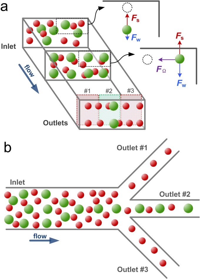
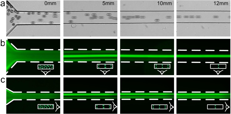
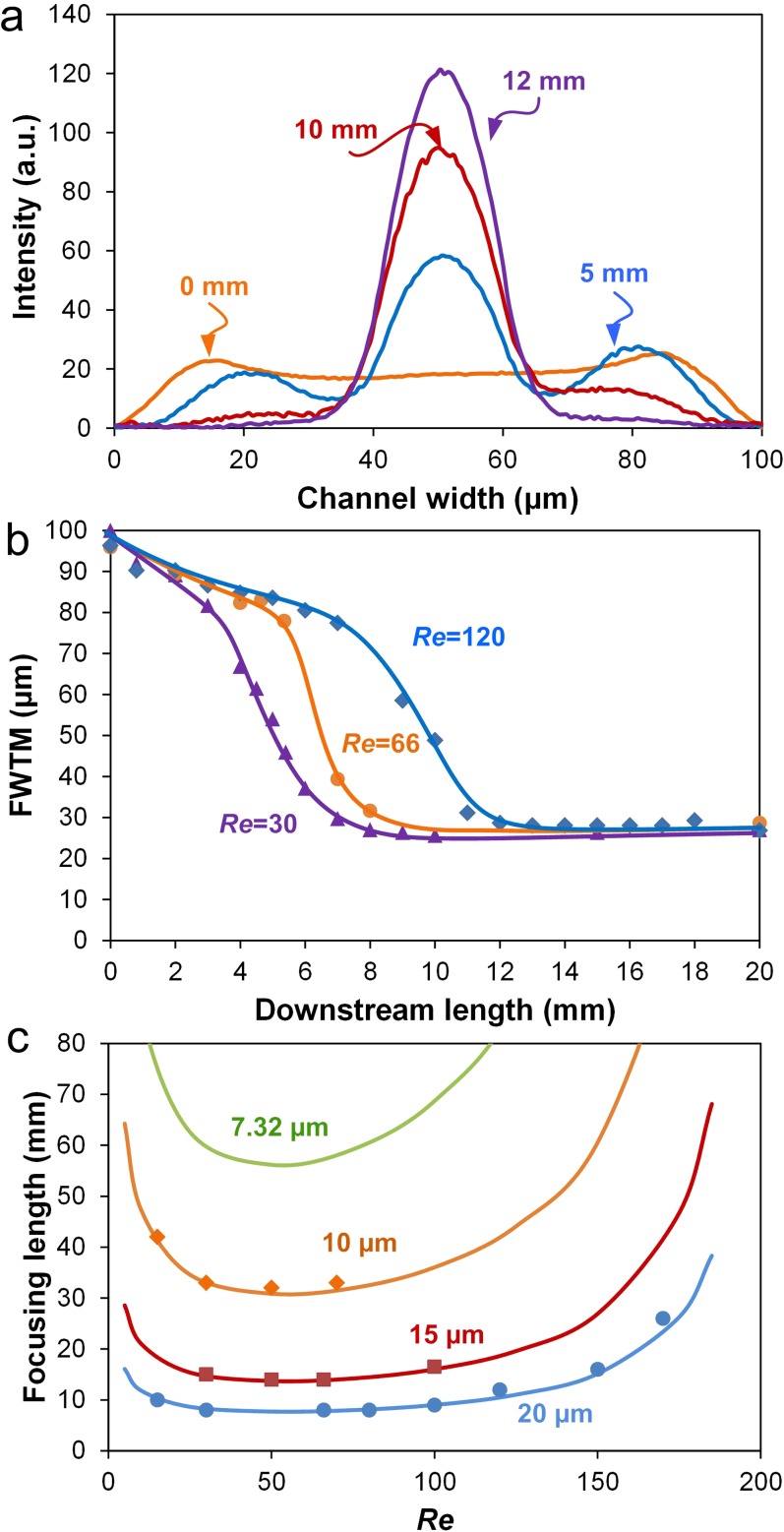
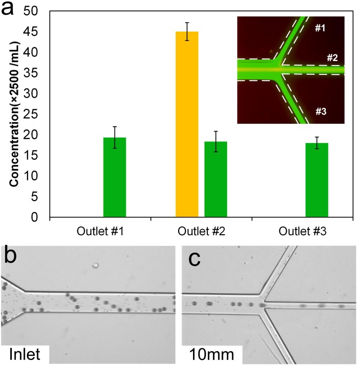
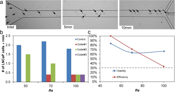
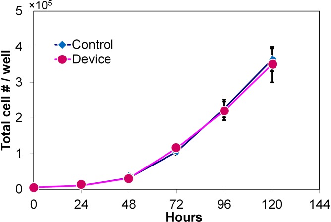
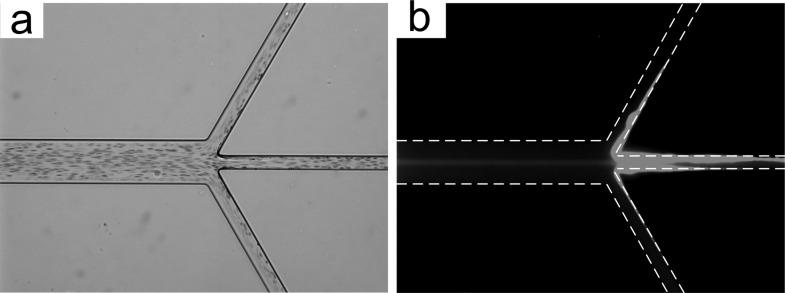
Cell filtration is a critical step in sample preparation in many bioapplications. Herein, we report on a simple, filter-free, microfluidic platform based on hydrodynamic inertial migration. Our approach builds on the concept of two-stage inertial migration which permits precise prediction of microparticle position within the microchannel. Our design manipulates equilibrium positions of larger microparticles by modulating rotation-induced lift force in a low aspect ratio microchannel. Here, we demonstrate filtration of microparticles with extreme efficiency (>99%). Using multiple prostate cell lines (LNCaP and human prostate epithelial tumor cells), we show filtration from spiked blood, with 3-fold concentration and >83% viability. Results of a proliferation assay show normal cell division and suggest no negative effects on intrinsic properties. Considering the planar low-aspect-ratio structure and predictable focusing, we envision promising applications and easy integration with existing lab-on-a-chip systems.


Figures

FIG. 1.
FIG. 1.
Schematic (a) and device layout (b). Particles flowing in a low aspect-ratio microchannel first migrate to top and bottom walls undergoing a shear-induced lift force FS (stage 1); once the shear-induced lift force balances with a wall-induced lift force FW, particles further migrate toward the centers of horizontal walls experiencing a rotation-induced lift force FΩ (stage 2). Due to the strong size-dependent migration velocity, the larger particles complete the two-stage migration and equilibrate to the two long-face-centered positions much faster than the smaller particles. By setting the channel length to the focusing length of the larger particles, they can be filtered from the smaller particles through the central outlet.
FIG. 2.
FIG. 2.
Demonstration of two-position focusing at Re = 120 in a 100 μm × 50 μm microchannel. Topview of bright field (a) and corresponding fluorescent (b) images show the evolution of particle streams in a rectangular microchannel. (c) Corresponding images of sideview at progressive downstream positions. The polystyrene particles were 20 μm in diameter.
FIG. 3.
FIG. 3.
Measurements of focusing length. (a) Fluorescent intensity line scans across channel width at consecutive downstream positions. (b) FWTM of major peaks as a function of downstream length at various Re. (c) Focusing length as a function of Re. The prediction curves were calculated based on two-stage migration model. Solid symbols indicate experimental measurements. All data were obtained using polystyrene beads (10 μm, 15 μm, and 20 μm in diameter) in a 100 μm × 50 μm microchannel.
FIG. 4.
FIG. 4.
Demonstration of size-selective filtration at Re = 50. (a) Filtration of 20 μm diameter particles from a mixture with 7.32 μm diameter particles. Large particles (yellow stream in the inset) were collected through the central outlet, while smaller particles (green in the inset) were evenly distributed in all three outlets. Error bars represent standard deviations of three counts. (b) Bright field image of the inlet illustrating suspension of 20 μm particles spiked in blood. Small dots in the channel are blood cells. (c) Particles exiting through the central outlet and RBCs exiting through every outlet.
FIG. 5.
FIG. 5.
Validation of cell focusing in the microchannel. (a) Bright field images showing development of LNCaP cell train at different downstream positions at Re = 50. (b) Cells collected at each outlet at three Re. Control represents the cells collected without passing through the device. (c) Efficiency and viability data for a 10 mm long microchannel.
FIG. 6.
FIG. 6.
Proliferation results illustrating the LNCaP cell population that was filtered in the device vs. control population. The control sample consisted of cells that did not pass through the device. The error bars represent one standard deviation of independent three measurements.
FIG. 7.
FIG. 7.
Filtration of HPET cells from blood at Re = 50. (a) Brightfield image illustrating cells (mostly RBCs) in the microchannel. (b) Corresponding fluorescent image illustrating a stream of HPET cells in the middle of the channel.

Similar articles

Cited by

References

    1. Nilsson A., Petersson F., Jonsson H., and Laurell T., “ Acoustic control of suspended particles in micro fluidic chips,” Lab Chip 4, 131–135 (2004).10.1039/b313493h - DOI - PubMed
    1. Agarwal G. and Livermore C., “ Chip-based size-selective sorting of biological cells using high frequency acoustic excitation,” Lab Chip 11, 2204–2211 (2011).10.1039/c1lc20050j - DOI - PubMed
    1. Gou H.-L., Zhang X.-B., Bao N., Xu J.-J., Xia X.-H., and Chen H.-Y., “ Label-free electrical discrimination of cells at normal, apoptotic and necrotic status with a microfluidic device,” J. Chromatogr. A 1218, 5725–5729 (2011).10.1016/j.chroma.2011.06.102 - DOI - PubMed
    1. Gascoyne P. R., Noshari J., Anderson T. J., and Becker F. F., “ Isolation of rare cells from cell mixtures by dielectrophoresis,” Electrophoresis 30, 1388–1398 (2009).10.1002/elps.200800373 - DOI - PMC - PubMed
    1. Khoshmanesh K., Baratchi S., Tovar-Lopez F. J., Nahavandi S., Wlodkowic D., Mitchell A., and Kalantar-zadeh K., “ On-chip separation of Lactobacillus bacteria from yeasts using dielectrophoresis,” Microfluid. Nanofluid. 12, 597–606 (2012).10.1007/s10404-011-0900-8 - DOI

LinkOut - more resources