. 2014 Oct 7:4:858-71.
doi: 10.1016/j.fob.2014.09.007. eCollection 2014.

Structural and functional characterization of salmon STAT1, STAT2 and IRF9 homologs sheds light on interferon signaling in teleosts

Affiliations

Structural and functional characterization of salmon STAT1, STAT2 and IRF9 homologs sheds light on interferon signaling in teleosts

Mehrdad Sobhkhez et al. FEBS Open Bio. .

Abstract

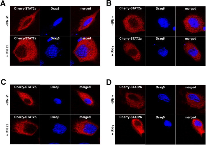
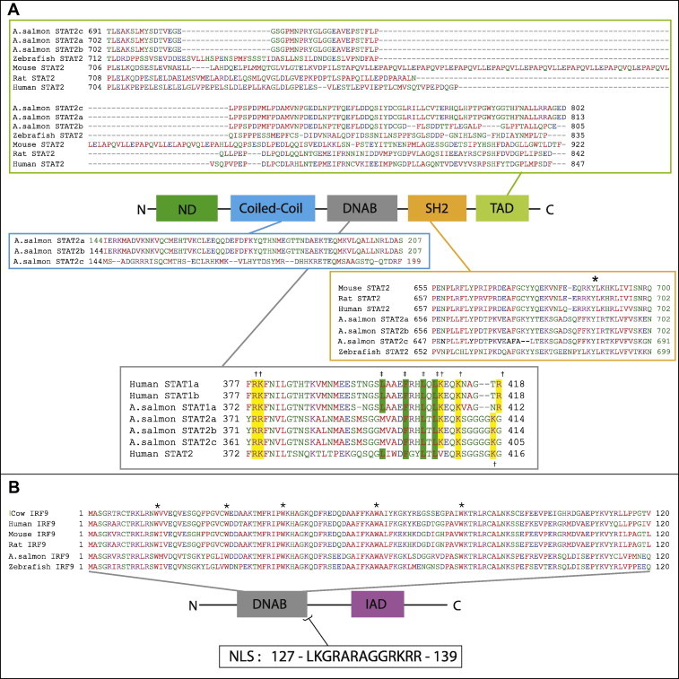
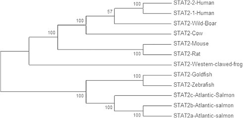
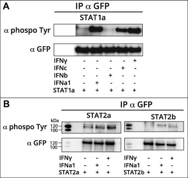
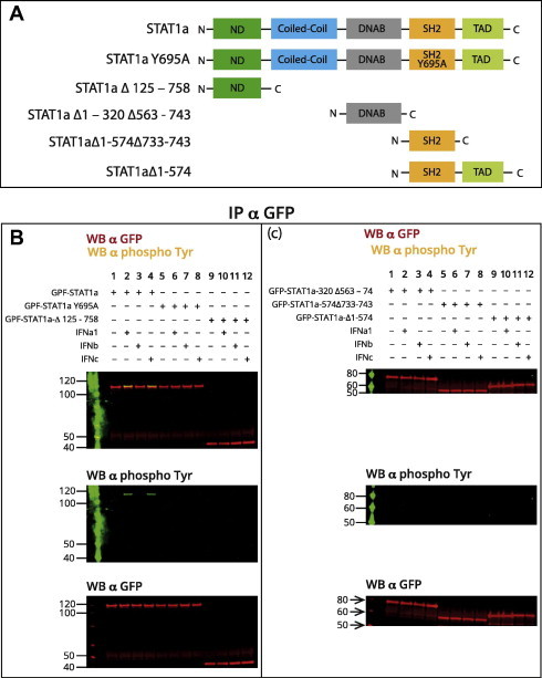
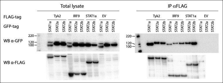
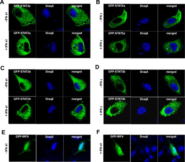
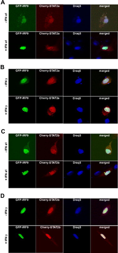
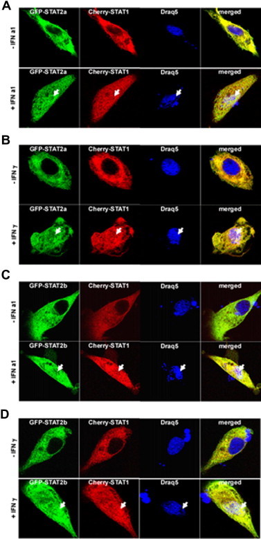
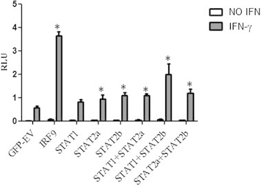
Mammalian IRF9 and STAT2, together with STAT1, form the ISGF3 transcription factor complex, which is critical for type I interferon (IFN)-induced signaling, while IFNγ stimulation is mediated by homodimeric STAT1 protein. Teleost fish are known to possess most JAK and STAT family members, however, description of their functional activity in lower vertebrates is still scarce. In the present study we have identified two different STAT2 homologs and one IRF9 homolog from Atlantic salmon (Salmo salar). Both proteins have domain-like structures with functional motifs that are similar to higher vertebrates, suggesting that they are orthologs to mammalian STAT2 and IRF9. The two identified salmon STAT2s, named STAT2a and STAT2b, showed high sequence identity but were divergent in their transactivation domain (TAD). Like STAT1, ectopically expressed STAT2a and b were shown to be tyrosine phosphorylated by type I IFNs and, interestingly, also by IFNγ. Microscopy analyses demonstrated that STAT2 co-localized with STAT1a in the cytoplasm of unstimulated cells, while IFNa1 and IFNγ stimulation seemed to favor their nuclear localization. Overexpression of STAT2a or STAT2b together with STAT1a activated a GAS-containing reporter gene construct in IFNγ-stimulated cells. The highest induction of GAS promoter activation was found in IFNγ-stimulated cells transfected with IRF9 alone. Taken together, these data suggest that salmon STAT2 and IRF9 may have a role in IFNγ-induced signaling and promote the expression of GAS-driven genes in bony fish. Since mammalian STAT2 is primarily an ISGF3 component and not involved in IFNγ signaling, our finding features a novel role for STAT2 in fish.

Keywords: Atlantic salmon; Fish; GAF, gamma interferon activation factor; IAD, IRF-association domain; IFN; IFN, type I interferon; IRF, interferon regulatory factor; IRF9; ISGF3, IFN-stimulated gene factor 3; ISRE, IFN-stimulated response element; NES, nuclear export signal; NLS, nuclear localization signal; STAT2; TAD, transcriptional activation domain.


Figures

Supplementary Fig. S1
Fig. 1
Fig. 1
Salmon STAT2 proteins harbor conserved domains and sequences. (A) The NCBI conserved domains database (CDD) and ClustalW alignment showed presence of conserved domains; N-terminal domain (ND), Coiled-coil, DNA binding domain (DBD), SH2 homology domain (SH2) and transcription activation domain (TAD). ClustalW alignment revealed high aa identity between the three identified salmon STAT2 sequences (STAT2a KJ155789, STAT2b KJ155790 and STAT2c FJ173070) and the highest divergence between STAT2c and STAT2a/STAT2b was observed in their Coiled-coil domain (aa 144–199/207). A conserved tyrosine residue (Y) which is a target of phosphorylation by the JAKs is marked with *. DNAB domain of STAT possess conserved aa essential for nuclear localization (marked yellow with †) and nuclear export (marked green with ‡). (B) DNAB domain of Atlantic salmon IRF9 has high sequence identity with IRF9 DNAB domains from other species. ClustalW alignment using the NCBI CDD of the deduced aa sequences of IRF9 DNAB domain from Atlantic salmon (NP_001167190), human (NP_006075), cow (AAI02048), mouse (AAH05435), rat (NP_001012041 XP_224190) and zebrafish (AAH81591). Asterisk (*): indicate conserved tryptophan (W) residues. (For interpretation of the references to colour in this figure legend, the reader is referred to the web version of this article.)
Fig. 2
Fig. 2
A phylogenetic tree of STAT2 protein sequences in Atlantic salmon and selected vertebrate species. The phylogenetic tree was constructed by the neighbor-joining method in the MEGA 5 program and the bootstrap values were calculated with 500 replicates. STAT2 from following species are included: Cow, goldfish, zebrafish, human, mouse, rat, wild boar, western clawed frog, and the three STAT2 isoforms of Atlantic salmon. GenBank accession numbers of the sequences are listed in Table 1.
Fig. 3
Fig. 3
Relative gene expression of STAT2a/b and IRF9 in tissues from healthy Atlantic salmon The relative expression of STAT2a/b (A) and IRF9 (B) in the tissues heart, gill, intestine, spleen, posterior kidney (P. Kidney), head kidney (H. K), liver, skin, thymus and muscle were measured by qPCR and presented relative to EFlαβ by the formula (ctEFlαβ)/(ct of STAT2 or IRF9) Samples from 6 fish were analyzed and error bars represents the mean ±SD.
Fig. 4
Fig. 4
Atlantic salmon STAT1a, STAT2a and STAT2b are tyrosine phosphorylated upon IFN stimulation. (A) CHSE-214 cells were transfected with plasmids expressing GFP-tagged STAT1a and either were left unstimulated or were stimulated with 200 U/ml IFNa1, b and c and 500 ng/ml IFNγ for 30 min before harvest. (B) CHSE-214 cells were transfected with GFP-tagged STAT2a and STAT2b. The cells were either left unstimulated or they were stimulated with 200 U/ml IFNa1, and 500 ng/ml IFNγ for 30 min before harvest. IP was performed with anti-GFP antibody and tyrosine-phosphorylated proteins were detected by Western blot using anti-phosphotyrosine antibody. The membranes were stripped and re-probed with anti-GFP antibody to detect the total amount of immunoprecipitated proteins. These experiments were repeated 3 times with the same result (results not shown).
Fig. 5
Fig. 5
Salmon STAT1a is phosphorylated on a single site (Tyr 695) in the SH2-domain in type I IFN-stimulated cells and this phosphorylation requires expression of full-length STAT1a. (A) Constructs used for immunoprecipitations: full-length STAT1a, STAT1a Y695A and 4 different deletion mutants. (B) CHSE-214 cells were transfected with plasmids encoding different GFP-tagged STAT1a, STAT1a deletion mutants or point mutated STAT1a Y695A. The cells were harvested 72 h post transfection, and 30 min prior to harvest they were stimulated with 200 U/ml of IFNa1, b or c. Cell extracts were immunoprecipitated with anti-GFP antibody and tyrosine-phosphorylation was detected by immunoblotting using α-phospho-tyrosine antibody. The membrane was stripped and re-probed with α-GFP to detect the total amount of immunoprecipitated proteins.
Fig. 6
Fig. 6
STAT1a interacts with Tyk2 and IRF9, and STAT2a and STAT2b interact with Tyk2, IRF9 and STAT1a. HEK 293 cells co-transfected with plasmids expressing TYK2, IRF9 or STAT1a with FLAG-tag in combination with plasmids expressing either STAT2a or STAT2b with GFP-tag. The study also included plasmids expressing FLAG-tagged Tyk2 or IRF9 in combination with STAT1a with GFP-tag. The IP was performed with anti-FLAG M2 affinity gel and interacting partners were detected by Western blot using antibodies against GFP, membrane was stripped and treated with anti-FLAG antibody. An empty pDEST-3xFlag vector was included as control. All the proteins were shown to be expressed in total lysate.
Fig. 7
Fig. 7
STAT2a and STAT2b localize in the cytoplasm, whereas IRF9 reside in the nucleus TO cells. (A) GFP-STAT2a non-stimulated (upper panel) and stimulated with IFNa1 (lower panel). (B) GFP-STAT2a non-stimulated (upper panel) and stimulated with IFNγ (lower panel). (C) GFP-STAT2b non-stimulated (upper panel) and stimulated with IFNa1 (lower panel). (D) GFP-STAT2b non-stimulated (upper panel) and stimulated with IFNγ (lower panel). (E) GFP-IRF9 non-stimulated and (F) stimulated with IFNa1. Cultured TO cells was transfected with the indicated constructs for 48 h. Subsequently, the cells were stimulated either with IFNa1 or IFNγ before fixation and counterstaining with Draq5 DNA dye to visualize nuclei and nucleic acids. Laser scanning confocal microscopy was done using Zeiss LSM 510 and high-magnification image analysis was done using a Zeiss LSM image browser.
Fig. 8
Fig. 8
IRF9 alters the sub-cellular localization of STAT2a and STAT2b into nuclei of TO cells, independent of type I or type II IFN stimulation. (A) GFP-IRF9 and Cherry-STAT2a non-stimulated (upper panel) and stimulated with IFN a1 (lower panel). (B) GFP-IRF9 and Cherry-STAT2a non-stimulated (upper panel) and stimulated with IFNγ (lower panel). (C) GFP-IRF9 and Cherry-STAT2b non-stimulated (upper panel) and stimulated with IFN a1 (lower panel). (D) GFP-IRF9 and Cherry-STAT2b non-stimulated (upper panel) and stimulated with IFNγ (lower panel). Cultured TO cells were co-transfected with GFP-IRF9 together with Cherry-STAT2a or Cherry-STAT2b for forty-eight hours. Subsequently, the cells were either stimulated with IFNa1 or IFNγ before fixation and counterstaining with Draq5 DNA dye to visualize nuclei and nucleic acids. Laser scanning confocal microscopy was done using Zeiss LSM 510 and high-magnification image analysis was done using a Zeiss LSM image browser.
Fig. 9
Fig. 9
Sub-cellular localization of STAT2a and STAT2b together with STAT1a in salmon cells stimulated with type I or type II IFN. (A) GFP-STAT2a and Cherry-STAT1a non-stimulated (upper panel) and stimulated with IFNa1 (lower panel). (B) GFP-STAT2a and Cherry-STAT1a non-stimulated (upper panel) and stimulated with IFNγ (lower panel). (C) GFP-STAT2b and Cherry-STAT1a non-stimulated (upper panel) and stimulated with IFN a1 (lower panel). (D) GFP-STAT2b and Cherry-STAT1a non-stimulated (upper panel) and stimulated with IFNγ (lower panel). Cultured TO cells was co-transfected with the indicated constructs for forty-eight hours. Subsequently, the cells were either stimulated with IFNa1 or IFNγ before fixation and counterstaining with Draq5 DNA dye to visualize nuclei and nucleic acids. Laser scanning confocal microscopy was done using Zeiss LSM 510 and high-magnification image analysis was done using a Zeiss LSM image browser.
Fig. 10
Fig. 10
Expression kinetics of STAT2a, STAT2b and IRF9 in IFN-stimulated TO cells. TO cells were treated with 200 U/ml of IFNa1, IFNb and IFNc, and 100 ng/ml IFNγ for 4, 24 and 48 h and untreated cells were used as controls. The mRNA expression levels of STAT2 (A), IRF9 (B) and Mx (C) were measured by qPCR and transcripts levels were normalized against EF1αβ. The results are presented as fold increase relative to untreated cells in triplicate samples and are representative of two independent experiments.
Fig. 11
Fig. 11
IRF9, STAT2a and STAT2b stimulate GAS promoter activity upon IFNγ stimulation. CHSE-214 cells transiently transfected with STAT1a, STAT2a, STAT2b, RF9 alone or in combination with each other and GFP-EV (empty vector) was used as control. Transfectants were left non-stimulated or were stimulated with 500 ng/ml of IFNγ 24 h prior to reporter gene assay analysis. Firefly luciferase activity was normalized with Renilla luciferase levels (n = 5) and the data are presented as relative luciferase units (RLU). The results are representative of two independent experiments. Represents values where difference compared to EV control is statistically significant.

References

    1. Stark G.R., Darnell J.E., Jr. The JAK–STAT pathway at twenty. Immunity. 2012;36:503–514. - PMC - PubMed
    1. Rawlings J.S., Rosler K.M., Harrison D.A. The JAK/STAT signaling pathway. J. Cell Sci. 2004;117:1281–1283. - PubMed
    1. Platanias L.C., Uddin S., Colamonici O.R. Tyrosine phosphorylation of the alpha and beta subunits of the type I interferon receptor. Interferon-beta selectively induces tyrosine phosphorylation of an alpha subunit-associated protein. J. Biol. Chem. 1994;269:17761–17764. - PubMed
    1. Colamonici O., Yan H., Domanski P., Handa R., Smalley D. Direct binding to and tyrosine phosphorylation of the alpha subunit of the type I interferon receptor by p135tyk2 tyrosine kinase. Mol. Cell. Biol. 1994;14:8133–8142. - PMC - PubMed
    1. Colamonici O.R., Uyttendaele H., Domanski P., Yan H., Krolewski J.J. P135tyk2, an interferon-alpha-activated tyrosine kinase, is physically associated with an interferon-alpha receptor. J. Biol. Chem. 1994;269:3518–3522. - PubMed

LinkOut - more resources