

Arterial spin labelling reveals prolonged arterial arrival time in idiopathic Parkinson's disease
- PMID: 25379411
- PMCID: PMC4215519
- DOI: 10.1016/j.nicl.2014.07.014
Arterial spin labelling reveals prolonged arterial arrival time in idiopathic Parkinson's disease
Abstract
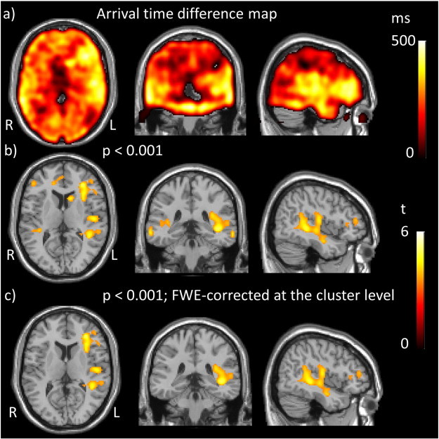
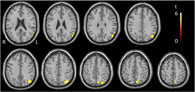
Idiopathic Parkinson's disease (IPD) is the second most common neurodegenerative disease, yet effective disease modifying treatments are still lacking. Neurodegeneration involves multiple interacting pathological pathways. The extent to which neurovascular mechanisms are involved is not well defined in IPD. We aimed to determine whether novel magnetic resonance imaging (MRI) techniques, including arterial spin labelling (ASL) quantification of cerebral perfusion, can reveal altered neurovascular status (NVS) in IPD. Fourteen participants with IPD (mean ± SD age 65.1 ± 5.9 years) and 14 age and cardiovascular risk factor matched control participants (mean ± SD age 64.6 ± 4.2 years) underwent a 3T MRI scan protocol. ASL images were collected before, during and after a 6 minute hypercapnic challenge. FLAIR images were used to determine white matter lesion score. Quantitative images of cerebral blood flow (CBF) and arterial arrival time (AAT) were calculated from the ASL data both at rest and during hypercapnia. Cerebrovascular reactivity (CVR) images were calculated, depicting the change in CBF and AAT relative to the change in end-tidal CO2. A significant (p = 0.005) increase in whole brain averaged baseline AAT was observed in IPD participants (mean ± SD age 1532 ± 138 ms) compared to controls (mean ± SD age 1335 ± 165 ms). Voxel-wise analysis revealed this to be widespread across the brain. However, there were no statistically significant differences in white matter lesion score, CBF, or CVR between patients and controls. Regional CBF, but not AAT, in the IPD group was found to correlate positively with Montreal cognitive assessment (MoCA) scores. These findings provide further evidence of alterations in NVS in IPD.
Keywords: 3T, 3 Tesla; AAT, arterial arrival time; AD, Alzheimer’s disease; ASL, arterial spin labelling; Arterial arrival time; Arterial spin labelling; CBF, cerebral blood flow; CO2, carbon dioxide; CV, cerebrovascular; CVD, cerebrovascular disease; CVR, cerebrovascular reactivity; CVRAAT, cerebrovascular reactivity measures of arterial arrival time; CVRCBF, cerebrovascular reactivity measures of cerebral blood flow; Cerebral blood flow; Cerebrovascular reactivity; DS, digit span; DSST, digit symbol substitution test; DWMH, deep white matter hyperintensity; EPI, echo planar imaging; ETCO2, end-tidal carbon dioxide; FAS, (verbal) fluency assessment scale; FLAIR, fluid attenuation inversion recovery; FWE, family-wise error; HAM-D, Hamilton depression rating scale; IPD, idiopathic Parkinson's disease; Idiopathic Parkinson's disease; L-dopa, levodopa; LARS, Lille apathy rating scale; LEDD, levodopa equivalent daily dose; MCI, mild cognitive impairment; MRI, magnetic resonance imaging; MoCA; MoCA, Montreal cognitive assessment; NPI, neuropsychiatric inventory; NVU, Neurovascular unit; O2−, oxygen; PET, positron emission tomography; PIGD, Postural instability and gait disorder; PL, parietal lobe; PVH, periventricular hyperintensity; ROI, region of interest; SPECT, single positron emission computed tomography; SPM, statistical parametric mapping; STAR, signal targeting with alternating radiofrequency; TD, tremor dominant; TE, echo time; TI, inversion time; TL, temporal lobe; TMT-B, trail making test B; TR, repetition time; UKPDS BB, United Kingdom Parkinson's Disease Society Brain Bank; UPDRS, Unified Parkinson's disease Rating Scale; WAIS-R, Wechsler adult intelligence scale-revised; WML, white matter lesion; fMRI, functional magnetic resonance imaging.
Figures
References
Publication types
MeSH terms
Substances
LinkOut - more resources
Full Text Sources
Other Literature Sources
Medical
Research Materials
Miscellaneous
