Randomized Controlled Trial
. 2014 Sep 16:6:252-61.
doi: 10.1016/j.nicl.2014.09.007. eCollection 2014.

Resting state connectivity correlates with drug and placebo response in fibromyalgia patients

Affiliations
Randomized Controlled Trial

Resting state connectivity correlates with drug and placebo response in fibromyalgia patients

T Schmidt-Wilcke et al. Neuroimage Clin. .

Abstract

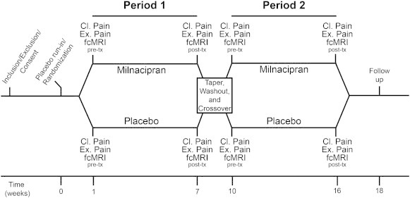
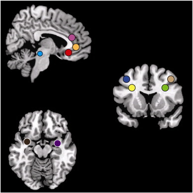
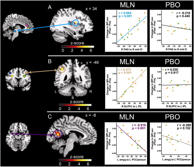
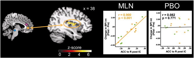
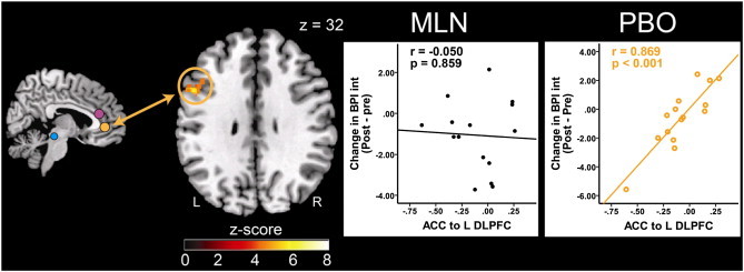
Fibromyalgia is a chronic pain syndrome characterized by widespread pain, fatigue, and memory and mood disturbances. Despite advances in our understanding of the underlying pathophysiology, treatment is often challenging. New research indicates that changes in functional connectivity between brain regions, as can be measured by magnetic resonance imaging (fcMRI) of the resting state, may underlie the pathogenesis of this and other chronic pain states. As such, this parameter may be able to be used to monitor changes in brain function associated with pharmacological treatment, and might also be able to predict treatment response. We performed a resting state fcMRI trial using a randomized, placebo-controlled, cross-over design to investigate mechanisms of action of milnacipran (MLN), a selective serotonin and norepinephrine reuptake inhibitor (SNRI), in fibromyalgia patients. Our aim was to identify functional connectivity patterns at baseline that would differentially predict treatment response to MLN as compared to placebo. Since preclinical studies of MLN suggest that this medication works by augmenting antinociceptive processes, we specifically investigated brain regions known to be involved in pain inhibition. 15 fibromyalgia patients completed the study, consisting of 6 weeks of drug and placebo intake (order counterbalanced) with an interspersed 2 week wash out period. As a main finding we report that reductions in clinical pain scores during MLN were associated with decreased functional connectivity between pro-nociceptive regions and antinociceptive pain regions at baseline, specifically between the rostral part of the anterior cingulate cortex (ACC) and the insular cortex (IC), as well as between the periaqueductal gray (PAG) and the IC: patients with lower preexisting functional connectivity had the greatest reduction in clinical pain. This pattern was not observed for the placebo period. However a more robust placebo response was associated with lower baseline functional connectivity between the ACC and the dorsolateral prefrontal cortex. This study indicates that ACC-IC connectivity might play a role in the mechanism of action of MLN, and perhaps more importantly fcMRI might be a useful tool to predict pharmacological treatment response.

Keywords: 5-HT, serotonin; ACC, anterior cingulate cortex; BPI, brief pain inventory; CNS, central nervous system; Chronic pain; DLPFC, dorsolateral prefrontal cortex; DMN, default mode network; FEW, family wise error; Fibromyalgia; Functional connectivity; IC, insular cortex; IPL, inferior parietal lobule; MCC, mid-cingulate cortex; MLN, milnacipran; NE, norepinephrine; PAG, periaqueductal gray; PCC, posterior cingulate cortex; QST, quantitative sensory testing; SNRI; SNRI, selective serotonin and norepinephrine reuptake inhibitor; SPM, statistical parametric mapping; TMS, transcranial magnetic stimulation; fMRI; fMRI, functional magnetic resonance imaging; fcMRI, functional connectivity magnetic resonance imaging; rs-fc, resting state functional connectivity.


Figures

Fig. 1
Fig. 1
Overview of study design. Subjects underwent a baseline visit where they were assessed for study criteria. Those that met inclusion/exclusion criteria were consented and randomized into the milnacipran or placebo treatment arm. Both clinical pain and experimental pain were assessed at each neuroimaging visit. Prior to their first neuroimaging session, subjects began a 1 week placebo run-in period. Patients that entered the milnacipran period first were dose escalated over 2 weeks to a stable dose of 200 mg/day, and maintained this fixed daily dose for 4 weeks. Subjects randomized to placebo for the first period took matching placebo pills for 6 weeks. After the 6 total weeks for this period, subjects underwent a post-treatment neuroimaging session. Following the completion of period one, all subjects underwent a 1 week taper and 2 weeks of placebo washout. After the washout period, participants crossed over to the other study drug for the second period. MRI = magnetic resonance imaging; tx = treatment.
Fig. 2
Fig. 2
Region of interest seeds included in the rs-fc analysis. Fig. 2 displays a priori seed regions included in the resting state functional connectivity analysis. Three anterior cingulate cortex spheres with radii of 5 mm included the ventral ACC (x = −3, y = 32, z = 19), the pre-genual ACC (x = 0, y = 40, z = 0), subgenual ACC (x = −3, y = 32, z = –8), two periaqueductal gray spherical seeds with 4 mm radii (left: x = 6, y = −26, z = –14; right: x = −6, y = −26, z = –14), four dorsolateral prefrontal cortex spherical seeds of 5 mm radii (left medial-inferior: x = −30, y = 15, z = 29; left lateral-superior: x = −40, y = 12, z = 44; right medial-inferior: x = 30, y = 15, z = 29; right lateral-superior: x = 40, y = 12, z = 44), and two amygdala spherical seeds of 5 mm radii (left: x = −32, y = 2, z = –16; right: x = 32, y = 2, z = 16).
Fig. 3
Fig. 3
Pre-MLN resting state functional connectivity predicts pain severity reductions in response to MLN treatment. Fig. 3 displays pre-milnacipran (MLN) treatment connectivity as a predictor for clinical response to MLN. Results displayed contain seed-to-target connectivity (seed regions displayed on the left) and plots of significant regressions for the MLN treatment arm (A. Mid-IC; B. DLPFC; C. PCC/precuneus), and corresponding statistics for the placebo treatment period. Orange dots represent patients that had PBO treatment first, and MLN treatment second. Green dots represent patients that received MLN treatment first, and PBO second. ACC = anterior cingulate cortex, amyg = amygdala, BPI Sev = Brief Pain Inventory severity scores, DLPFC = dorsolateral prefrontal cortex, IC = insular cortex, IPL = inferior parietal lobule, L = left, MLN = milnacipran, PAG = periaqueductal gray, PBO = placebo, PCC = posterior cingulate cortex, pre-cun = precuneus, R = right.
Fig. 4
Fig. 4
Pre-MLN resting state functional connectivity predicts decrease in pain interference reduction in response to MLN treatment. Fig. 4 displays pre-milnacipran (MLN) treatment connectivity as a predictor for pain response to MLN. Results displayed contain seed-to-target connectivity (seed regions displayed on left) and plots of significant regressions for the MLN treatment arm and corresponding statistics for the placebo treatment period. ACC = anterior cingulate cortex, BPI Int = Brief Pain Inventory interference scores, IC = insular cortex, IPL = inferior parietal lobule, L = left, MLN = milnacipran, PBO = placebo, R = right.
Fig. 5
Fig. 5
Pre-placebo resting state functional connectivity predicts clinical pain reduction in response to placebo treatment. Fig. 5 displays pre-placebo (PBO) treatment connectivity as a predictor for response to PBO pills. Results displayed contain seed-to-target connectivity (seed regions displayed on left) and plots of significant regressions for the PBO treatment arm, and corresponding statistics for the MLN treatment period. ACC = anterior cingulate cortex, BPI Int = Brief Pain Inventory interference scores, DLPFC = dorsolateral prefrontal cortex, L = left, MLN = milnacipran, PBO = placebo, R = right.

References

    1. Assumpção A., Cavalcante A.B., Capela C.E., Sauer J.F., Chalot S.D., Pereira C.A. Prevalence of fibromyalgia in a low socioeconomic status population. BMC Musculoskeletal Disorders. 2009;10:64. 19505321 - PMC - PubMed
    1. Beckmann C.F., DeLuca M., Devlin J.T., Smith S.M. Investigations into resting-state connectivity using independent component analysis. Philosophical Transactions of the Royal Society B: Biological Sciences. 2005;360(1457):1001–1013. - PMC - PubMed
    1. Bernardy K., Klose P., Busch A.J., Choy E.H., Häuser W. Cognitive behavioural therapies for fibromyalgia. Cochrane Database of Systematic Reviews. 2013;9:CD009796. 24018611 - PMC - PubMed
    1. Bingel U. [Mechanisms of endogenous pain modulation illustrated by placebo analgesia: functional imaging findings] Schmerz (Berlin, Germany) 2010;24(2):122–129. 20376600 - PubMed
    1. Bingel U., Schoell E., Herken W., Büchel C., May A. Habituation to painful stimulation involves the antinociceptive system. Pain. 2007;131(1–2):21–30. 17258858 - PubMed

Publication types