

Brain volume estimation from post-mortem newborn and fetal MRI
- PMID: 25379457
- PMCID: PMC4218943
- DOI: 10.1016/j.nicl.2014.10.007
Brain volume estimation from post-mortem newborn and fetal MRI
Abstract
Objective: Minimally invasive autopsy using post-mortem magnetic resonance imaging (MRI) is a valid alternative to conventional autopsy in fetuses and infants. Estimation of brain weight is an integral part of autopsy, but manual segmentation of organ volumes on MRI is labor intensive and prone to errors, therefore unsuitable for routine clinical practice. In this paper we aim to show that volumetric measurements of the post-mortem fetal and neonatal brain can be accurately estimated using semi-automatic techniques and a high correlation can be found with the weights measured from conventional autopsy results.
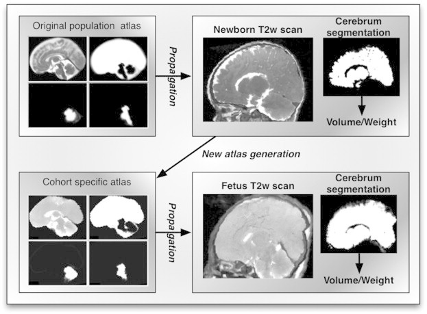
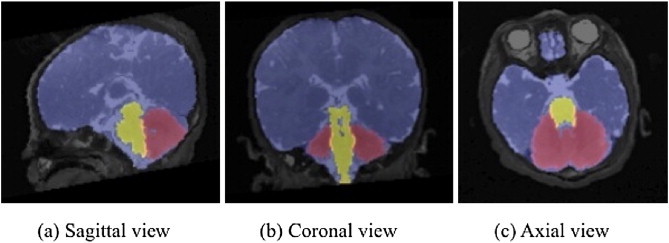
Methods: The brains of 17 newborn subjects, part of Magnetic Resonance Imaging Autopsy Study (MaRIAS), were segmented from post-mortem MR images into cerebrum, cerebellum and brainstem using a publicly available neonate brain atlas and semi-automatic segmentation algorithm. The results of the segmentation were averaged to create a new atlas, which was then used for the automated atlas-based segmentation of 17 MaRIAS fetus subjects. As validation, we manually segmented the MR images from 8 subjects of each cohort and compared them with the automatic ones. The semi-automatic estimation of cerebrum weight was compared with the results of the conventional autopsy.
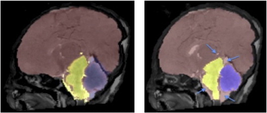
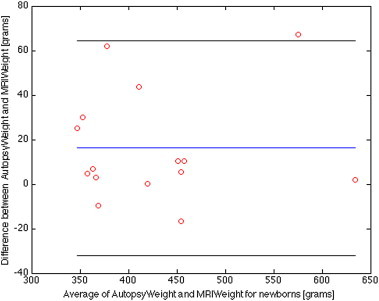
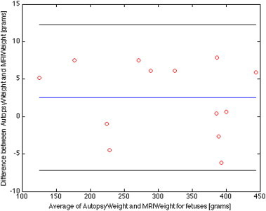
Results: The Dice overlaps between the manual and automatic segmentations are 0.991 and 0.992 for cerebrum, 0.873 and 0.888 for cerebellum and 0.819 and 0.815 for brainstem, for newborns and fetuses, respectively. Excellent agreement was obtained between the estimated MR weights and autopsy gold standard ones: mean absolute difference of 5 g and 2% maximum error for the fetus cohort and mean absolute difference of 20 g and 11% maximum error for the newborn one.
Conclusions: The high correlation between the obtained segmentation and autopsy weights strengthens the idea of using post-mortem MRI as an alternative for conventional autopsy of the brain.
Keywords: Autopsy; Brain volumes; CI, confidence interval; CSF, cerebrospinal fluid; Cerebrum; EM, expectation maximization; Fetus; GA, gestational age; GW, gestational weeks; MRI, magnetic resonance imaging; MaRIAS, Magnetic Resonance Imaging Autopsy Study; Newborn; Post-mortem MRI.
Figures
References
-
- Ashburner J. Computational Neuroanatomy. University College London; London: 2000.
-
- Breeze A.C., Cross J.J., Hackett G.A., Jessop F.A., Joubert I., Lomas D.J. Use of a confidence scale in reporting postmortem fetal magnetic resonance imaging. Ultrasound in Obstetrics & Gynecology: the Official Journal of the International Society of Ultrasound in Obstetrics and Gynecology. 2006;28(7):918–924. 17124693 - PubMed
Publication types
MeSH terms
Grants and funding
LinkOut - more resources
Full Text Sources
Other Literature Sources
Medical
Research Materials
