Congenital muscular torticollis concurrent with sagittal synostosis: a case report
- PMID: 25379504
- PMCID: PMC4221403
- DOI: 10.5535/arm.2014.38.5.712
Congenital muscular torticollis concurrent with sagittal synostosis: a case report
Abstract
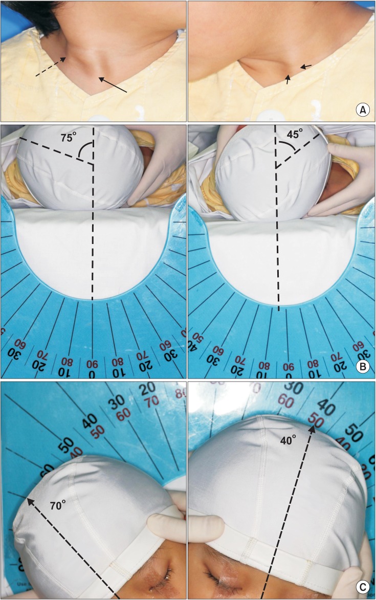
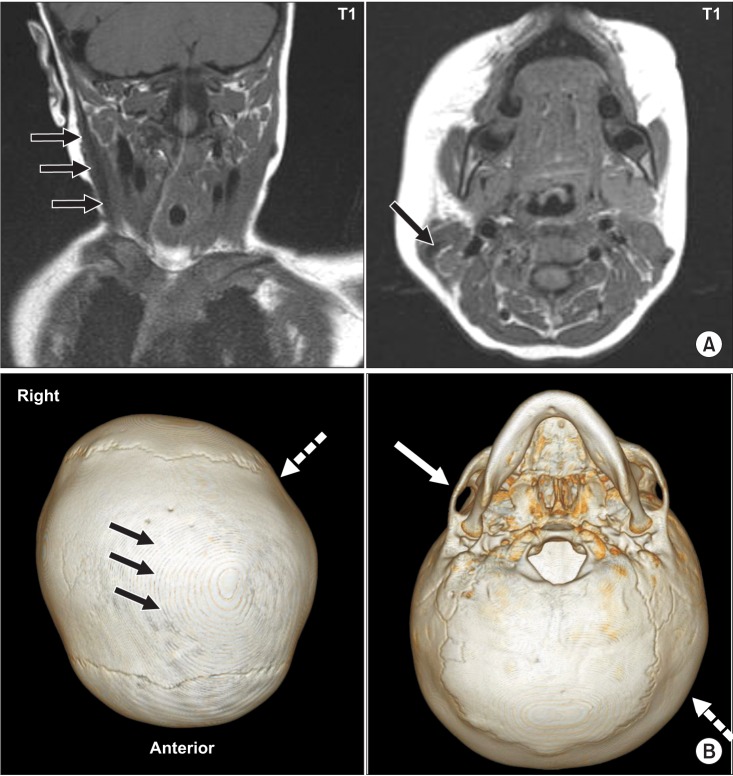
Congenital muscular torticollis (CMT) and craniosynostosis are diseases that cause plagiocephaly and craniofacial asymmetry in children. In our literature review, we did not find any report of concurrent manifestation of CMT and craniosynostosis. A 41-month-old boy visited our hospital with left torticollis, right laterocollis, and craniofacial asymmetry as the main findings. During clinical examination, prominent right sternocleidomastoid muscle and limited range of motion of the neck were noted, and right CMT was confirmed by magnetic resonance imaging of the neck. Three-dimensional computed tomography of the skull, which was conducted due to the unusual appearance of the skull with a large head circumference, mild brachycephaly, as well as left plagiocephaly, revealed premature closure of the sagittal suture. Thus, we report the first case that showed concurrence of CMT and sagittal synostosis. We recommend that concurrently manifested craniosynostosis needs to be examined if the subject with CMT displays unusual craniofacial asymmetry to a greater extent than deformational plagiocephaly.
Keywords: Craniosynostosis; Plagiocephaly; Torticollis.
Conflict of interest statement
No potential conflict of interest relevant to this article was reported.
Figures
Similar articles
-
Concurrence of Congenital Muscular Torticollis and Congenital Torticollis Due to Other Anomalies: Two Case Reports.Front Pediatr. 2021 Oct 27;9:709616. doi: 10.3389/fped.2021.709616. eCollection 2021. Front Pediatr. 2021. PMID: 34778123 Free PMC article.
-
Description of Mandibular Improvements in a Series of Infants With Congenital Muscular Torticollis and Deformational Plagiocephaly Treated With Physical Therapy.Cleft Palate Craniofac J. 2018 Oct;55(9):1282-1288. doi: 10.1177/1055665618763374. Epub 2018 Jul 10. Cleft Palate Craniofac J. 2018. PMID: 29989836
-
The role of congenital muscular torticollis in the development of deformational plagiocephaly.Plast Reconstr Surg. 2009 Feb;123(2):643-652. doi: 10.1097/PRS.0b013e318196b9be. Plast Reconstr Surg. 2009. PMID: 19182625
-
Deformational plagiocephaly associated with ocular torticollis: a clinical study and literature review.J Craniofac Surg. 2007 Mar;18(2):399-405. doi: 10.1097/SCS.0b013e3180341ca6. J Craniofac Surg. 2007. PMID: 17414292 Review.
-
Craniosynostosis.Am Fam Physician. 2004 Jun 15;69(12):2863-70. Am Fam Physician. 2004. PMID: 15222651 Review.
References
-
- Looman WS, Flannery AB. Evidence-based care of the child with deformational plagiocephaly, Part I: assessment and diagnosis. J Pediatr Health Care. 2012;26:242–250. - PubMed
-
- Badve CA, K MM, Iyer RS, Ishak GE, Khanna PC. Craniosynostosis: imaging review and primer on computed tomography. Pediatr Radiol. 2013;43:728–742. - PubMed
-
- Bendon CL, Sheerin FB, Wall SA, Johnson D. The relationship between scaphocephaly at the skull vault and skull base in sagittal synostosis. J Craniomaxillofac Surg. 2014;42:245–249. - PubMed
-
- Raco A, Raimondi AJ, De Ponte FS, Brunelli A, Bristot R, Bottini DJ, et al. Congenital torticollis in association with craniosynostosis. Childs Nerv Syst. 1999;15:163–169. - PubMed
LinkOut - more resources
Full Text Sources
Other Literature Sources