. 2014 Dec;11(6):551-9.
doi: 10.1089/zeb.2014.1024.

The tumor suppressor rpl36 restrains KRAS(G12V)-induced pancreatic cancer

Affiliations

The tumor suppressor rpl36 restrains KRAS(G12V)-induced pancreatic cancer

Elayne Provost et al. Zebrafish. 2014 Dec.

Abstract

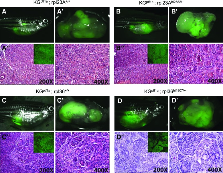
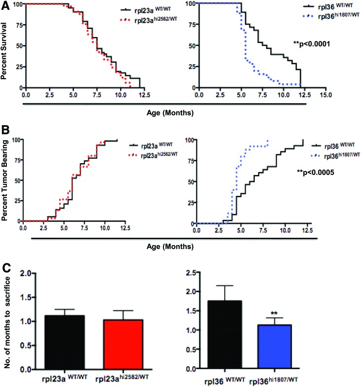
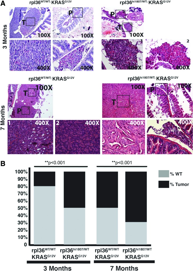
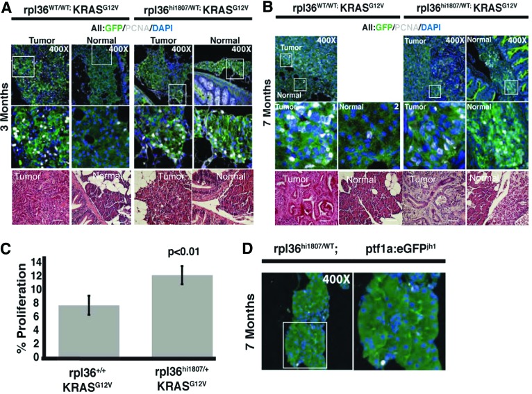
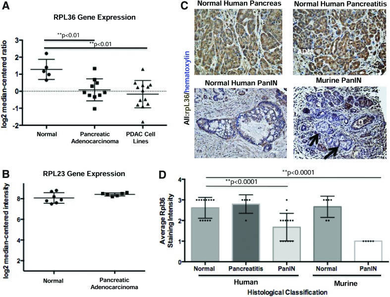
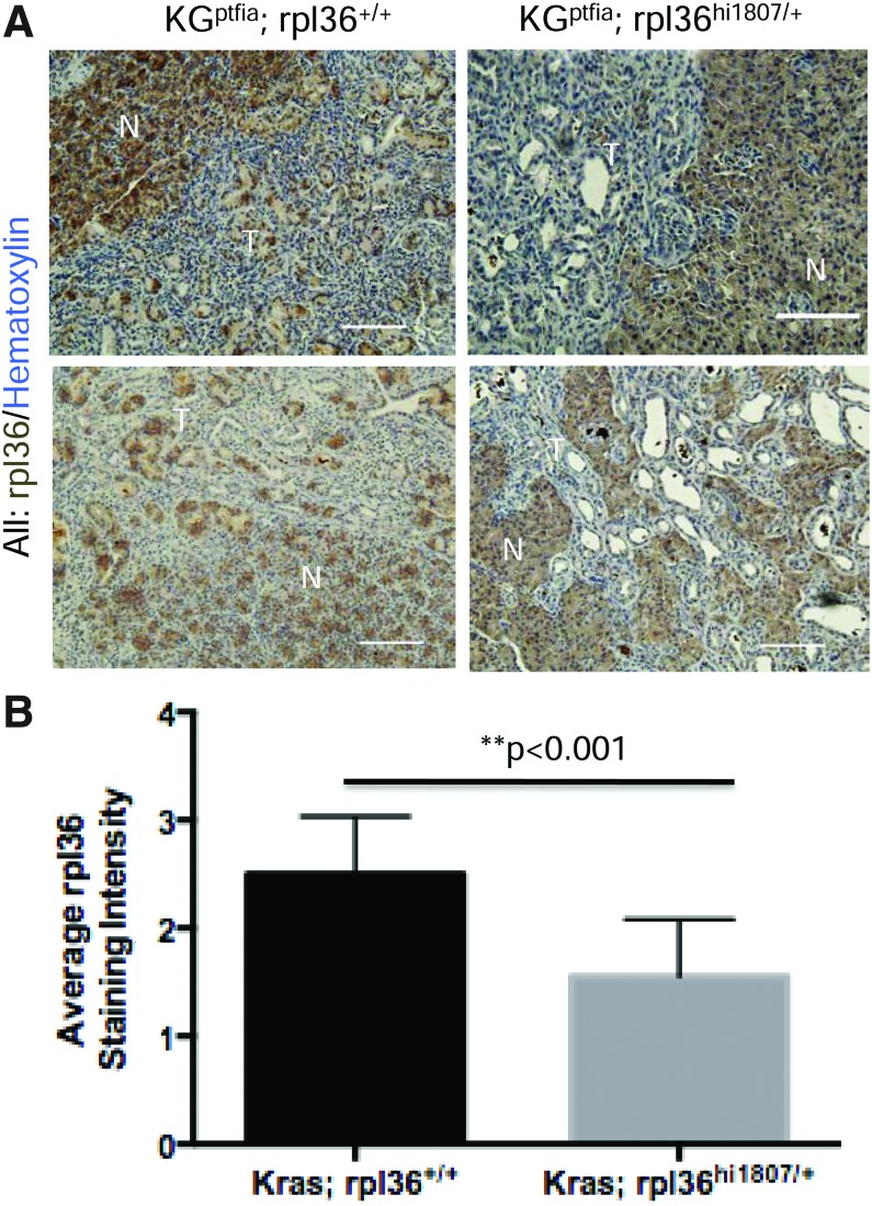
Ribosomal proteins are known to be required for proper assembly of mature ribosomes. Recent studies indicate an additional role for ribosomal proteins as candidate tumor suppressor genes. Pancreatic acinar cells, recently identified as effective cells of origin for pancreatic adenocarcinoma, display especially high-level expression of multiple ribosomal proteins. We, therefore, functionally interrogated the ability of two ribosomal proteins, rpl36 and rpl23a, to alter the response to oncogenic Kras in pancreatic acinar cells using a newly established model of zebrafish pancreatic cancer. These studies reveal that rpl36, but not rpl23a, acts as a haploinsufficient tumor suppressor, as manifested by more rapid tumor progression and decreased survival in rpl36(hi1807/+);ptf1a:gal4VP16(Tg);UAS:GFP-KRAS(G12V) fish compared with their rpl36(+/+);ptf1a:gal4VP16;UAS:GFP-KRAS(G12V) siblings. These results suggest that rpl36 may function as an effective tumor suppressor during pancreatic tumorigenesis.


Figures

<b>FIG. 1.</b>
FIG. 1.
Pancreatic tumor histology in the setting of haploinsufficiency for either rpl23a or rpl36. Representative images of 7-month-old rpl23aWT/WT; ptf1a:gal4-VP16; UAS:GFP-KRASG12V (A), rpl23ahi2582/WT; ptf1a:gal4-VP16; UAS:GFP-KRASG12V (B), rpl36WT/WT; ptf1a:gal4-VP16; UAS:GFP-KRASG12V (C) and rpl36hi1807/WT; ptf1a:gal4-VP16; UAS:GFP-KRASG12V fish (D). For each genotype, upper left images depict fish with distended abdomen and transcutaneous eGFP fluorescence; upper right images depict excised abdominal viscera confirming eGFP+pancreatic tumors. Lower panels show representative histology of mixed acinar/ductal tumors. Insets confirm expression of GFP-KRASG12V fusion protein in malignant tumor epithelium as assessed by antibody staining for GFP (representative of n=96 tumors analyzed). Color images available online at www.liebertpub.com/zeb
<b>FIG. 2.</b>
FIG. 2.
Haploinsufficiency for rpl36, but not rpl23a, accelerates pancreatic tumorigenesis in ptf1a:Gal4-VP16;UAS:GFP-KRASG12V transgenic zebrafish. (A) Age-dependent tumor incidence, (B) overall survival, and (C) time from tumor diagnosis to death for ptf1a:Gal4-VP16; UAS:GFP-KRASG12V (KGptf1a) fish with (red; n=34) and without (black; n=67) heterozygous rpl23a mutations and with (blue; n=26) and without (black; n=29) heterozygous rpl36 mutations. **p<0.01. Haploinsufficiency for rpl23a has no effect on tumor onset, tumor progression, or overall survival. In contrast, haploinsufficiency for rpl36 accelerates tumor onset and tumor progression, and is associated with diminished overall survival. Color images available online at www.liebertpub.com/zeb
<b>FIG. 3.</b>
FIG. 3.
Loss of rpl36 increases the percentage of transgenic zebrafish with pancreatic tumors. (A) Histological analysis of tumors derived from KGptf1a; rpl36+/+ or KGptf1a; rpl36hi1807/+ zebrafish at 3 or 7 months of age at the time of sacrifice. Histology of dissected viscera with both non-neoplastic pancreas (P) and tumor tissue (T) for both zebrafish genotypes is represented. No differences in tumor histology are observed in the presence or absence of rpl36 haploinsufficiency. (B) The percentage of fish with normal histological pancreas (gray) or tumor (black) in 3- and 7-month-old rpl36hi1807/WT;ptf1a:gal4-VP16;UAS:GFP-KRASG12V fish and their rpl36WT/WT;ptf1a:gal4-VP16;UAS:GFP-KRASG12V control siblings (**p<0.001). Color images available online at www.liebertpub.com/zeb
<b>FIG. 4.</b>
FIG. 4.
Loss of rpl36 results in an increase in proliferation in ptf1a:gal4-VP16;UAS:GFP-KRASG12V pancreas. (A, B), determination of cell proliferation using PCNA labeling (white nuclei) in tumor and normal pancreatic tissue harvested from control rpl36WT/WT;ptf1a:gal4-VP16;UAS:GFP-KRASG12V and mutant rpl36hi1807/WT;ptf1a:gal4-VP16;UAS:GFP-KRASG12V fish at 3 months (A) and 7 months (B) of age. Increased proliferation was detected only in tumor tissue harvested from rpl36WT/WT fish, and not in adjacent normal tissue. In contrast, increased proliferation is seen in both tumor and adjacent normal tissue in ptf1a:gal4-VP16;UAS:GFP-KRASG12V fish with heterozygous rpl36hi1807 mutation (C). Quantification of proliferative index of normal pancreatic tissue from control rpl36WT/WT;ptf1a:gal4-VP16;UAS:GFP-KRASG12V and mutant rpl36hi1807/WT;ptf1a:gal4-VP16;UAS:GFP-KRASG12V 3 month-old adult zebrafish. Students t-test, **p<0.01. (D) PCNA labeling of rpl36hi1807/WT zebrafish showing rpl36 haploinsufficiency does not increase proliferation in the absence of oncogenic KRAS. Color images available online at www.liebertpub.com/zeb
<b>FIG. 5.</b>
FIG. 5.
Immunohistochemical analysis of RPL36 in human and murine pancreatic tissue. (A, B) Representative gene expression data for RPL36 and RPL23a. (ONCOMINE). Microarray data confirm that RPL36, but not RPL23a, transcript abundance is decreased in bulk human pancreatic ductal adenocarcinoma specimens and in human PDAC cell lines relative to normal pancreas (data extracted from Iacobuzio-Donahue et al., 2003). (C) Immunohistochemistry for RPL36. Analysis was performed on normal human pancreatic tissue, human chronic pancreatitis, and human PanIN. Immunohistochemistry was also performed on murine PanINs from Mist1:CreERT2;LSL-KrasG12D PanIN-bearing mice (n=3). Note high-level expression in normal pancreatic duct (D) but absent expression in PanIN lesions (arrows). (D) Graphical representation of average RPL36 staining intensity (1=light staining; 3=dark staining) as a function of pathological classification (n=minimum of 10 patient samples per group). RPL36 staining intensity was significantly decreased in human and murine PanIN relative to normal pancreatic tissue (**p<0.0001). Color images available online at www.liebertpub.com/zeb
<b>FIG. 6.</b>
FIG. 6.
Immunohistochemical analysis of zebrafish tumors reveals a significant reduction of rpl36 expression in KGptf1a;rpl36hi1807/+ tumors compared with KGptf1a;rpl36+/+ tumors. (A) IHC staining for rpl36 in a panel of tumor sections derived from either KGptf1a; rpl36hi1807/+ or KGptf1a; rpl36+/+ zebrafish (n=8 per group). (B) Quantification of staining intensity (1=light brown staining, 3=dark brown staining) in the tumor sections analyzed. Tumors occurring in fish with heterozygous loss of rpl36 displayed less intense labeling for rpl36. Color images available online at www.liebertpub.com/zeb

References

    1. Feldmann G, Beaty R, Hruban RH, Maitra A. Molecular genetics of pancreatic intraepithelial neoplasia. J Hepatobiliary Pancreat Surg 2007;14:224–232 - PMC - PubMed
    1. Corbo V, Tortora G, Scarpa A. Molecular pathology of pancreatic cancer: from bench-to-bedside translation. Curr Drug Targets 13:744–752 - PMC - PubMed
    1. Provost E, et al. . Ribosomal biogenesis genes play an essential and p53-independent role in zebrafish pancreas development. Development 139:3232–3241 - PMC - PubMed
    1. Amsterdam A, et al. . A large-scale insertional mutagenesis screen in zebrafish. Genes Dev 1999;13:2713–2724 - PMC - PubMed
    1. Amsterdam A, et al. . Many ribosomal protein genes are cancer genes in zebrafish. PLoS Biol 2004;2:E139. - PMC - PubMed

Publication types

LinkOut - more resources