. 2015 Jan;156(1):255-67.
doi: 10.1210/en.2014-1675.

Expression and distribution of glucagon-like peptide-1 receptor mRNA, protein and binding in the male nonhuman primate (Macaca mulatta) brain

Affiliations

Expression and distribution of glucagon-like peptide-1 receptor mRNA, protein and binding in the male nonhuman primate (Macaca mulatta) brain

Kristy M Heppner et al. Endocrinology. 2015 Jan.

Abstract

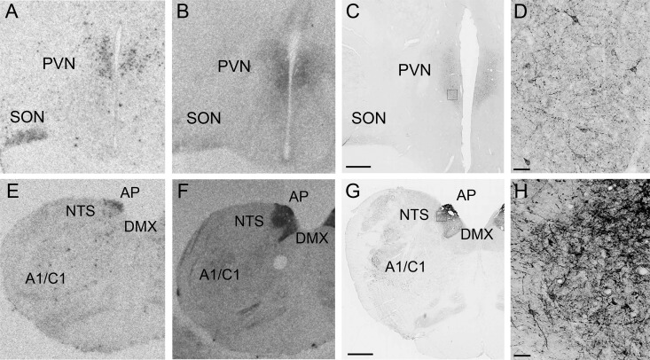
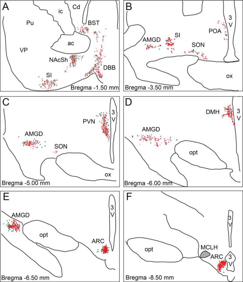
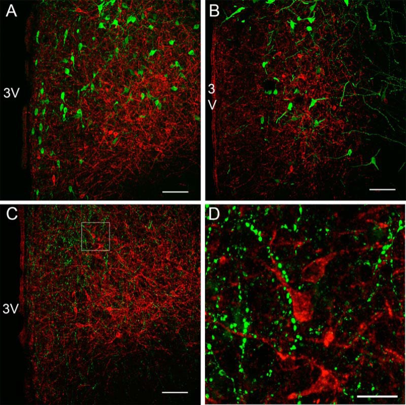
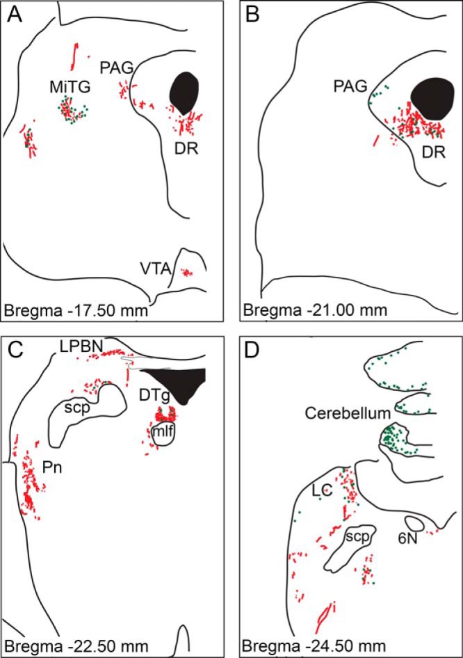
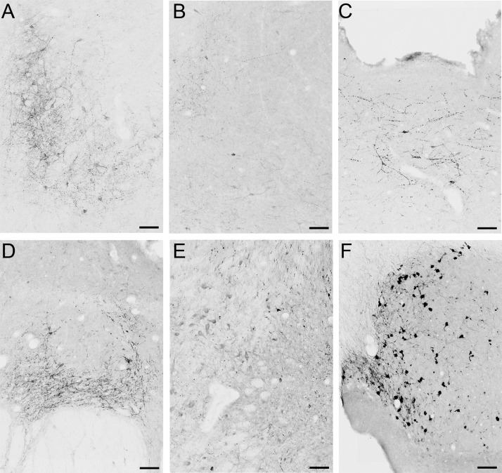
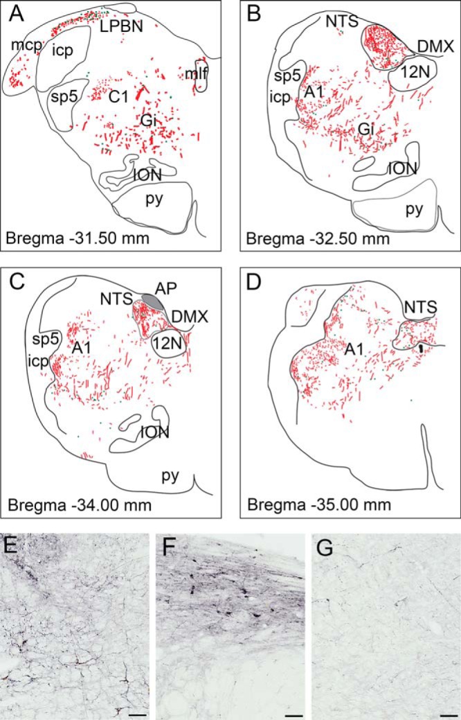
Glucagon-like peptide-1 (GLP-1) is released from endocrine L-cells lining the gut in response to food ingestion. However, GLP-1 is also produced in the nucleus of the solitary tract, where it acts as an anorectic neurotransmitter and key regulator of many autonomic and neuroendocrine functions. The expression and projections of GLP-1-producing neurons is highly conserved between rodent and primate brain, although a few key differences have been identified. The GLP-1 receptor (GLP-1R) has been mapped in the rodent brain, but no studies have described the distribution of GLP-1Rs in the nonhuman primate central nervous system. Here, we characterized the distribution of GLP-1R mRNA and protein in the adult macaque brain using in situ hybridization, radioligand receptor autoradiography, and immunohistochemistry with a primate specific GLP-1R antibody. Immunohistochemistry demonstrated that the GLP-1R is localized to cell bodies and fiber terminals in a very selective distribution throughout the brain. Consistent with the functional role of the GLP-1R system, we find the highest concentration of GLP-1R-immunoreactivity present in select hypothalamic and brainstem regions that regulate feeding, including the paraventricular and arcuate hypothalamic nuclei, as well as the area postrema, nucleus of the solitary tract, and dorsal motor nucleus of the vagus. Together, our data demonstrate that GLP-1R distribution is highly conserved between rodent and primate, although a few key species differences were identified, including the amygdala, where GLP-1R expression is much higher in primate than in rodent.


Figures

Figure 1.
Figure 1.. Confirmation of GLP-1R antibody specificity using ISH and ISLB.
GLP-1R distribution using ISH (A and E), ISLB (B and F), and IHC (C and D and G and H) in the hypothalamus at the level of the PVN (A–D) and AP/NTS (E–H). D, High-power magnification of C. H, High-power magnification of G. Scale bars, 1000 μm (C and G) and 50 μm (D and H).
Figure 2.
Figure 2.. Schematic representation of GLP-1R-ir distribution in the NHP forebrain.
Depiction of GLP-1R-ir cell bodies (green dots) and GLP-1R-ir fibers (red lines) in forebrain sections arranged from rostral (A) to caudal (F). Gray shaded area in the magnocellular nucleus of the lateral hypothalamus (MCLH) indicates dense fiber network. 3V, third ventricle; ac, anterior commissure; BST, bed nucleus of the stria terminalis; Cd, caudate; DBB, diagonal band of Broca; DMH, dorsomedial nucleus of the hypothalamus; ic, internal capsule; NAcSh, nucleus accumbens shell; opt, optic tract; ox, optic chiasm; Pu, putamen; SI, substantia innominata; SON, supraoptic nucleus; VP, ventral pallidum.
Figure 3.
Figure 3.. Photomicrographs of GLP-1R-ir in the NHP forebrain.
GLP-1R-ir was detected in the diagonal band of Broca (DBB) (A), nucleus accumbens shell (NAcSh) (B), POA (C), DMH (D), AMGD (E, image at the level of the ARC), and ARC (F). 3V, third ventricle; DMH, dorsomedial nucleus of the hypothalamus. Scale bars, 100 μm.
Figure 4.
Figure 4.. Distribution of GLP-1R immunofluorescence with anorexogenic neuronal populations in the NHP PVN.
Confocal microscopic images showing GLP-1R-ir (red) with oxytocin-ir (A; green), AVP-ir (B; green) or CART-ir (C; green). D, High-power magnification of C. 3V, third ventricle. Scale bars, 100 μm (A–C) 25 μm (D).
Figure 5.
Figure 5.. Schematic representation of GLP-1R-ir distribution in the NHP midbrain.
Depiction of GLP-1R-ir cell bodies (green dots) and GLP-1R-ir fibers (red lines) in midbrain sections arranged from rostral (A) to caudal (D). 6N, abducens nucleus; MiTG, microcellular tegmental nucleus; mlf, medial longitudinal fasciculus; Pn, pontine reticular nucleus; scp, superior cerebellar peduncle.
Figure 6.
Figure 6.. Photomicrographs of GLP-1R-ir in the NHP midbrain.
GLP-1R-ir was detected in the microcellular tegmental nucleus (MiTG) (A), PAG (B), DR (C), DTg (D), LC (E), and cerebellum (F). Scale bars, 100 μm.
Figure 7.
Figure 7.. Schematic representation and photomicrographs of GLP-1R-ir distribution in the NHP hindbrain.
Depiction of GLP-1R-ir cell bodies (green dots) and GLP-1R-ir fibers (red lines) in hindbrain sections arranged from rostral (A) to caudal (D). Gray shaded area in the AP indicates dense fiber network. Photomicrographs of GLP-1R-ir are shown in the A1 area (E), LPBN (F), and Gi (G). 12N, hypoglossal nucleus; A1, noradrenergic cell group; C1, adrenergic cell group; icp, inferior cerebellar peduncle; ION, inferior olivary nucleus; mcp, middle cerebellar peduncle; mlf, medial longitudinal fasciculus; py, pyramidal tract; sp5, spinal trigeminal tract. Scale bars, 100 μm.

References

    1. Kreymann B, Williams G, Ghatei MA, Bloom SR. Glucagon-like peptide-1 7-36: a physiological incretin in man. Lancet. 1987;2(8571):1300–1304. - PubMed
    1. Mojsov S, Weir GC, Habener JF. Insulinotropin: glucagon-like peptide I (7-37) co-encoded in the glucagon gene is a potent stimulator of insulin release in the perfused rat pancreas. J Clin Invest. 1987;79(2):616–619. - PMC - PubMed
    1. Willard FS, Sloop KW. Physiology and emerging biochemistry of the glucagon-like peptide-1 receptor. Exp Diabetes Res. 2012;2012:470851. - PMC - PubMed
    1. Edwards KL, Stapleton M, Weis J, Irons BK. An update in incretin-based therapy: a focus on glucagon-like peptide-1 receptor agonists. Diabetes Technol Ther. 2012;14(10):951–967. - PubMed
    1. Merchenthaler I, Lane M, Shughrue P. Distribution of pre-pro-glucagon and glucagon-like peptide-1 receptor messenger RNAs in the rat central nervous system. J Comp Neurol. 1999;403(2):261–280. - PubMed

Publication types