Review
. 2015 Jan;14(1):2-12.
doi: 10.1128/EC.00225-14. Epub 2014 Nov 7.

Targeting Toxoplasma tubules: tubulin, microtubules, and associated proteins in a human pathogen

Affiliations
Review

Targeting Toxoplasma tubules: tubulin, microtubules, and associated proteins in a human pathogen

Naomi Morrissette. Eukaryot Cell. 2015 Jan.

Abstract

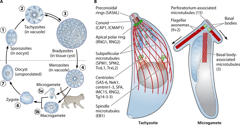
Toxoplasma gondii is an obligate intracellular parasite that causes serious opportunistic infections, birth defects, and blindness in humans. Microtubules are critically important components of diverse structures that are used throughout the Toxoplasma life cycle. As in other eukaryotes, spindle microtubules are required for chromosome segregation during replication. Additionally, a set of membrane-associated microtubules is essential for the elongated shape of invasive "zoites," and motility follows a spiral trajectory that reflects the path of these microtubules. Toxoplasma zoites also construct an intricate, tubulin-based apical structure, termed the conoid, which is important for host cell invasion and associates with proteins typically found in the flagellar apparatus. Last, microgametes specifically construct a microtubule-containing flagellar axoneme in order to fertilize macrogametes, permitting genetic recombination. The specialized roles of these microtubule populations are mediated by distinct sets of associated proteins. This review summarizes our current understanding of the role of tubulin, microtubule populations, and associated proteins in Toxoplasma; these components are used for both novel and broadly conserved processes that are essential for parasite survival.


Figures

FIG 1
FIG 1
(A) Organization of the microtubule cytoskeleton during the Toxoplasma life cycle. Infection of diverse vertebrate hosts occurs by ingestion of oocysts harboring sporozoites (1) or tissue cysts harboring bradyzoites (3). The released zoites infect and replicate in cells of the intestine to create tachyzoites (2), which disseminate throughout the host to infect diverse cell types. After development of an immune response to infection, tachyzoites differentiate into bradyzoites, a slow-growing form that persists for the life of the host organism. Bradyzoites can differentiate back into tachyzoites if host immunity wanes. In cats, tachyzoites and bradyzoites form merozoites (4), and these give rise to microgametes (5a) and macrogametes (5b). A transiently diploid zygote (6) is produced by fertilization, and becomes an unsporulated oocyst (7) after formation of an exterior cell wall. Oocysts are shed in feces and sporulate at ambient temperatures, creating two sporocysts, each containing four sporozoites (1). (B) A schematic of microtubule populations in tachyzoites and microgametes. (Left) The zoite stages have conserved microtubule structures which are best defined in tachyzoites. The spindle and subpellicular microtubule populations (red) associate with centrioles and the APR MTOCs (green). Defined components of these structures are noted. An unusual tubulin-based structure, the conoid, is located above the APR and can be withdrawn into it. There are two preconoidal rings above the conoid and two intraconoid microtubules within its circumference. (Right) Toxoplasma microgametes have two flagella, which originate at basal bodies in the apical region. Above these basal bodies is a pointed perforatorium structure which contains 15 short microtubules. A set of 5 peripheral microtubules extends from the basal body region toward the posterior with a longitudinal alignment. Due to the difficulty in obtaining the gamete stages, specific protein markers of these microtubules have not been identified. The images in panel B are based on previous figures in references , , and 139).
None

References

    1. Amos LA. 2011. What tubulin drugs tell us about microtubule structure and dynamics. Semin Cell Dev Biol 22:916–926. doi:10.1016/j.semcdb.2011.09.014. - DOI - PubMed
    1. Jordan A, Hadfield JA, Lawrence NJ, McGown AT. 1998. Tubulin as a target for anticancer drugs: agents which interact with the mitotic spindle. Med Res Rev 18:259–296. doi:10.1002/(SICI)1098-1128(199807)18:4<259::AID-MED3>3.0.CO;2-U. - DOI - PubMed
    1. Pellegrini F, Budman DR. 2005. Review: tubulin function, action of antitubulin drugs, and new drug development. Cancer Invest 23:264–273. doi:10.1081/CNV-200055970. - DOI - PubMed
    1. Lu Q, Luduena RF. 1994. In vitro analysis of microtubule assembly of isotypically pure tubulin dimers. Intrinsic differences in the assembly properties of alpha beta II, alpha beta III, and alpha beta IV tubulin dimers in the absence of microtubule-associated proteins. J Biol Chem 269:2041–2047. - PubMed
    1. Rezania V, Azarenko O, Jordan MA, Bolterauer H, Luduena RF, Huzil JT, Tuszynski JA. 2008. Microtubule assembly of isotypically purified tubulin and its mixtures. Biophys J 95:1993–2008. doi:10.1529/biophysj.108.132233. - DOI - PMC - PubMed

Publication types

MeSH terms