. 2015 Jan 2;290(1):544-55.
doi: 10.1074/jbc.M114.617555. Epub 2014 Nov 7.

Microseconds simulations reveal a new sodium-binding site and the mechanism of sodium-coupled substrate uptake by LeuT

Affiliations

Microseconds simulations reveal a new sodium-binding site and the mechanism of sodium-coupled substrate uptake by LeuT

Elia Zomot et al. J Biol Chem. .

Abstract

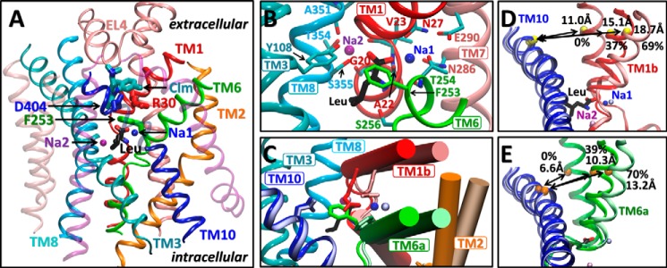
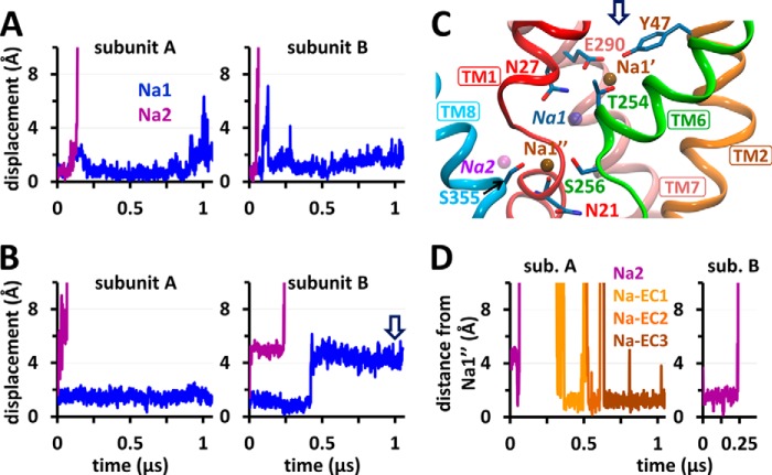
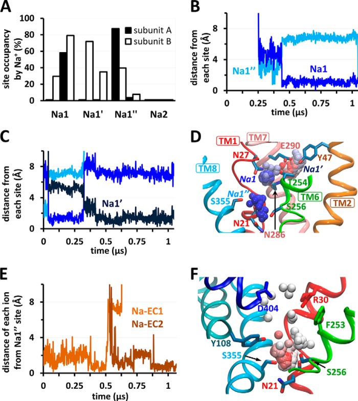
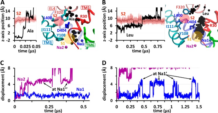
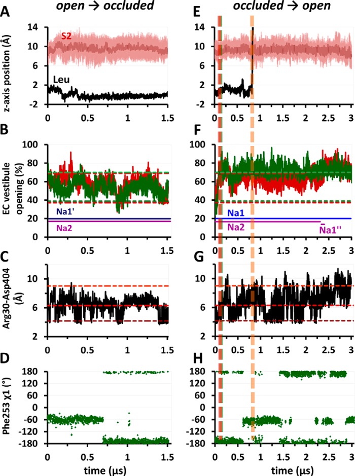
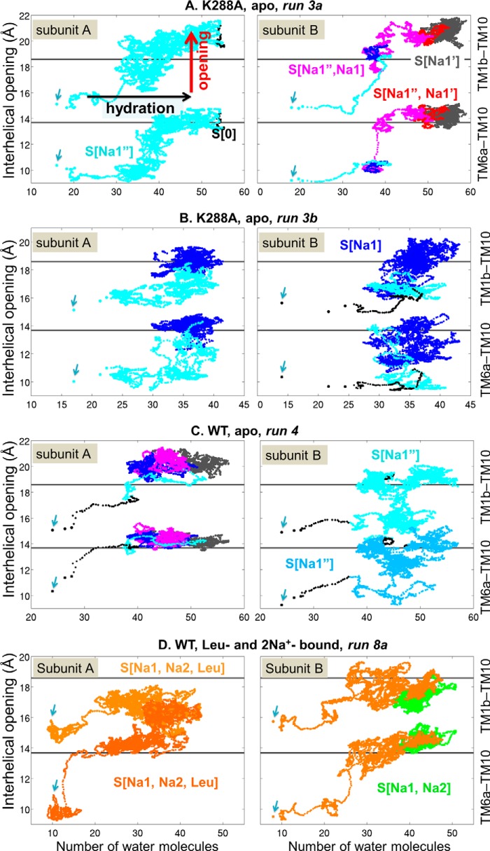
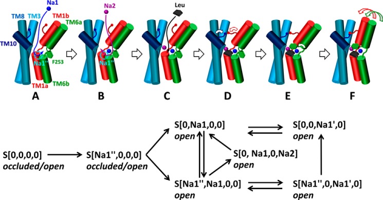
The bacterial sodium-coupled leucine/alanine transporter LeuT is broadly used as a model system for studying the transport mechanism of neurotransmitters because of its structural and functional homology to mammalian transporters such as serotonin, dopamine, or norepinephrine transporters, and because of the resolution of its structure in different states. Although the binding sites (S1 for substrate, and Na1 and Na2 for two co-transported sodium ions) have been resolved, we still lack a mechanistic understanding of coupled Na(+)- and substrate-binding events. We present here results from extensive (>20 μs) unbiased molecular dynamics simulations generated using the latest computing technology. Simulations show that sodium binds initially the Na1 site, but not Na2, and, consistently, sodium unbinding/escape to the extracellular (EC) region first takes place at Na2, succeeded by Na1. Na2 diffusion back to the EC medium requires prior dissociation of substrate from S1. Significantly, Na(+) binding (and unbinding) consistently involves a transient binding to a newly discovered site, Na1″, near S1, as an intermediate state. A robust sequence of substrate uptake events coupled to sodium bindings and translocations between those sites assisted by hydration emerges from the simulations: (i) bindings of a first Na(+) to Na1″, translocation to Na1, a second Na(+) to vacated Na1″ and then to Na2, and substrate to S1; (ii) rotation of Phe(253) aromatic group to seclude the substrate from the EC region; and (iii) concerted tilting of TM1b and TM6a toward TM3 and TM8 to close the EC vestibule.

Keywords: Biophysics; LeuT; Mechanism of Binding; Membrane Transport; NSS; Neurotransmitter Transport; Sodium Transport; Sodium-coupled; Transport.


Figures

FIGURE 1.
FIGURE 1.
Structure, binding sites, and conformational changes in LeuT. A, LeuT monomeric unit in OF-occluded form (PDB code 3F3E), viewed from the side. Leu (in CPK sticks; carbons in black) is shielded from the EC region by Phe253 side chain (green) on TM6. Na+ ions at sites Na1 and Na2 are shown in blue and purple, respectively. The secondary binding site (S2) binds larger noncompetitive inhibitors such as clomipramine (Clm, sticks, teal) (PDB code 2Q6H). B, a close up on the coordination of Leu and Na+ ions as viewed from the EC space. C, superposition of OF-occluded and -open (PDB code 3TT1) structures (darker and lighter colors, respectively), showing the motions in TM1b, 2 and 6a (cylinders), the rotation of Phe253 side chain, and the displacement of Arg30 (dark/light red) away from Asp404 (dark/light blue). D and E, changes in distances between Val33(C) and Asp401(Cα) (D, yellow spheres) and Ile245(C) and Ile410(Cα) (E, orange spheres), used as probes for TM1b–TM10 and TM6a–TM10 separations, respectively. The distances in the crystal structures used in this study are indicated, along with their relative openings with respect to the experimentally resolved inward facing occluded state (0%) and the maximal opening (100%) observed in simulations.
FIGURE 2.
FIGURE 2.
Sodium unbinding and binding events in the OF-open state of LeuT dimer in the absence of substrate and identification of a novel Na+-binding site Na1″. A and B, displacements of sodium ions originally located at Na1 and Na2 (blue and purple, respectively) away from their crystal positions, presented for subunits A and B in double mutant Y108F/K288A (A) and WT-LeuT (runs 1 and 2, respectively; Table 1) (B). C, a snapshot of subunit B of WT-LeuT (run 2) at ∼1 μs (arrow in B) shows the locations of two sites, Na1′ and Na1″, observed to stably bind sodium ions, and the vacated Na1 and Na2 sites (transparent blue and purple, respectively). The sodium ion at Na1′ (originally at Na1) is coordinated by Glu290 (TM7) and polar residues (sticks). Na+ at Na1″ is coordinated by Asn21, Ser256, Ser355, and Asn286 (omitted for clarity). D, high propensity of Na1″ site, 5–6 Å away from Na2, for binding Na+ ions, either on their way out to the EC environment (for Na+ ions originally bound to Na2; purple) or coming in from the EC environment (Na-EC1, Na-EC2, or Na-EC3, orange-brown). The data in D refer to run 2 (B). sub., subunit.
FIGURE 3.
FIGURE 3.
Time-dependent occupancy of sodium-binding sites Na1, Na1′, and Na1″ and sequential binding events as the OF-occluded K288A mutant transitions into OF-open state. A, in the absence of substrate and sodium ions (runs 3a and 3b), EC sodium ions enter the EC vestibule of K288A mutant and bind to the sites Na1, Na1′, and Na1″, but not to Na2, as shown by the percentage occupancies of these sites by Na+. B, the instantaneous position of an EC Na+ ion that enters subunit A (run 3b), using as metric its distances from the sites Na1″ (blue) and Na1 (dark blue). The EC Na+ ion first comes into close proximity of Na1″ and then binds Na1 where it settles for the remaining 0.6 μs of simulation. C, the behavior of another EC Na+ ion (subunit B; run 3a), which first recognizes and momentarily binds at Na1″ and then switches to Na1 (blue) and Na1′ (dark blue). D, the trajectory in C is illustrated where the spheres display the instantaneous positions of EC Na+ sampled at 1.25-ns intervals during 0 < t < 1 μs (color-coded by time from blue to red). E, stable binding of two EC Na+ ions (orange and brown) to Na1″ in subunit A (run 3a). F, the trajectory of the second EC Na+ ion (Na-EC2) in D is shown here for the time period 0.5 < t < 1 μs. On-pathway gating and/or Na+-coordinating residues are shown in sticks.
FIGURE 4.
FIGURE 4.
Substrate translocation to the EC environment in OF-open form and accompanying movements of sodium ions. A and B illustrate the translocation of Ala (run 5, subunit A) and Leu (run 6a, subunit A), respectively. The left panels show the instantaneous position of substrate along the z axis (perpendicular to membrane) (black curve), along with those of the center and upper/lower boundaries of the S2 site (dark pink and pink curves, respectively). The S1 site is at z = 0. The substrate moves “upwards” into the EC region. The right panels illustrate the successive positions (black spheres) of Ala or Leu (Cα-atoms), along the pathway to the EC region as viewed from the side sampled at 1.25ns. S1 and S2 sites are shown (yellow and pink circles, respectively). Residues that define the sites S1 (Tyr108; TM3) and S2 (Ile111 (TM3), Leu400 (TM10), and Phe320 (EL4)) and the EC gating residues Phe253 (green), Arg30 (red), and Asp404 (blue) are shown in sticks. The accompanying displacements of Na+ ions away from their original locations at Na1 and Na2 are shown in C and D, respectively. The arrows indicate the temporary bindings from Na2 to Na1″ (C) or from Na1 to Na1′ (D).
FIGURE 5.
FIGURE 5.
Gating and structural changes associated with binding and release of leucine in the OF state. Forward (A–D) and reverse (E–H) transitions between open and occluded forms, accompanying the respective binding or unbinding of leucine are shown. A and E, instantaneous position of Leu along the z axis (black) with respect to S1 site (at z = 0) and that of the S2 site (dark/light pink). B and F, the degree of opening of TM1b and 6a (red and green, respectively), as well as the occupancy of Na1, Na2, Na1′, or Na1″ sites by sodium ions initially at Na1 or Na2 (blue or purple, respectively) for each subunit. C and G, distance between Arg30:Cζ and Asp404:Cγ. The dashed lines in dark, medium, and light red indicate the crystal structure values for the direct, water-mediated, or no interactions (4.2, 6.3, or 9.0 Å, respectively) observed in IF-open, OF-occluded, and OF-open states. D and H, time evolution of Phe253 χ1 dihedral. The average values −67° and −164° correspond to the OF-open and -occluded crystal structures, respectively. E–H, vertical dashed lines indicate the times of TM1b opening (red), TM6a opening (green, overlaps with red line), and substrate unbinding from S1 (orange). The data are shown for subunit B in run 6a (A–D) and B in run 8a (E–H).
FIGURE 6.
FIGURE 6.
Coupling between the hydration of the EC vestibule and the transition from occluded to open OF state. The panels illustrate the trajectories of LeuT (WT or K288A), subunits A (left panels) and B (right panels), starting from occluded, minimally hydrated states (indicated by arrows). Each panel contains two sets of data points, displaying the respective interhelical distances TM1b–TM10 (based on Val33–Asp401 Cα-Cα distance) and TM6a–TM10 (Cα-Cα distance of Ile245–Ile410) (right ordinate) plotted as a function of the number of water molecules in the sodium/Leu binding pocket, defined as the region within 3 Å from ion/substrate coordinating residues as follows: (i) Na1″: Asn21, Ser256, and Ser355; (ii) Na1 and Na1′: Ala22, Asn27, Tyr47, Thr254, Asn286, and Glu290; (iii) Na2: Gly20, Val23, Ala351, Thr354, and Ser355; and (iv) S1: Ala22, Leu25, Gly26, Val104, Tyr108, Phe253, Thr354, Gly258, Ile359, Gly260, Ala261, and Ile262. The colors refer to different sodium/substrate-bound states, as labeled, e.g. S[0] (black), apo state; S[Na1″] (cyan), single Na+, bound at Na1″; S(Na1″, Na1) (magenta) two Na+ ions bound at Na1″ and Na1, etc. In C (right panel) and D (left panel), slightly different shades are used to distinguish the two trajectories.
FIGURE 7.
FIGURE 7.
Mechanism of sodium- and substrate-binding in the OF state of LeuT. A–E, in the OF-open LeuT, a sodium ion entering the EC vestibule from the EC region first binds to the Na1 site (A), followed by another at Na2 (B) (blue and purple spheres, respectively), with the entry path for both likely to be through Na1″ site (cyan circle). Leucine (black) then enters into the EC vestibule and binds to S1 (C), with little or no binding to S2. Although the EC vestibule is open (A–C), a water-mediated or direct interaction between Arg30 and Asp404 (blue and red sticks, respectively) can take place, which, together with the isomeric rotation of the aromatic ring of Phe253 (green hexagon), enables the EC gate closure (D). Closure of TM1b and 6a and formation of a stable salt bridge between Arg30 and Asp404 stabilize the substrate- and sodium-loaded occluded state (E). The lower panel provides a schematic description of the initial sodium binding events along with subsequent translocations observed in the simulations.

References

    1. Richerson G. B., Wu Y. (2004) Role of the GABA transporter in epilepsy. Adv. Exp. Med. Biol. 548, 76–91 - PubMed
    1. Andersen J., Kristensen A. S., Bang-Andersen B., Stromgaard K. (2009) Recent advances in the understanding of the interaction of antidepressant drugs with serotonin and norepinephrine transporters. Chem. Commun. (Camb.) 25, 3677–3692 - PubMed
    1. Shaffer P. L., Goehring A., Shankaranarayanan A., Gouaux E. (2009) Structure and mechanism of a Na+-independent amino acid transporter. Science 325, 1010–1014 - PMC - PubMed
    1. Clausen R. P., Madsen K., Larsson O. M., Frølund B., Krogsgaard-Larsen P., Schousboe A. (2006) Structure-activity relationship and pharmacology of γ-aminobutyric acid (GABA) transport inhibitors. Adv. Pharmacol. 54, 265–284 - PubMed
    1. Hahn M. K., Blakely R. D. (2002) Monoamine transporter gene structure and polymorphisms in relation to psychiatric and other complex disorders. Pharmacogenomics J. 2, 217–235 - PubMed

Publication types

MeSH terms

Substances

LinkOut - more resources