. 2014 Dec 26;289(52):36249-62.
doi: 10.1074/jbc.M114.613901. Epub 2014 Nov 7.

Insights into the specificity of lysine acetyltransferases

Affiliations

Insights into the specificity of lysine acetyltransferases

Alex C Tucker et al. J Biol Chem. .

Abstract

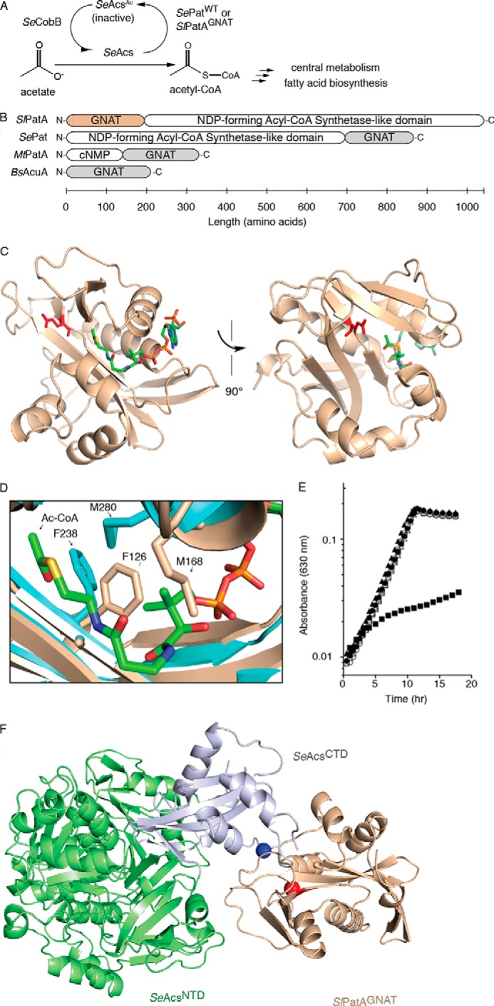
Reversible lysine acetylation by protein acetyltransferases is a conserved regulatory mechanism that controls diverse cellular pathways. Gcn5-related N-acetyltransferases (GNATs), named after their founding member, are found in all domains of life. GNATs are known for their role as histone acetyltransferases, but non-histone bacterial protein acetytransferases have been identified. Only structures of GNAT complexes with short histone peptide substrates are available in databases. Given the biological importance of this modification and the abundance of lysine in polypeptides, how specificity is attained for larger protein substrates is central to understanding acetyl-lysine-regulated networks. Here we report the structure of a GNAT in complex with a globular protein substrate solved to 1.9 Å. GNAT binds the protein substrate with extensive surface interactions distinct from those reported for GNAT-peptide complexes. Our data reveal determinants needed for the recognition of a protein substrate and provide insight into the specificity of GNATs.

Keywords: Acetyl Coenzyme A (Acetyl-CoA); Bacterial Metabolism; Enzyme Inactivation; Histone Acetylase; Post-translational Modification (PTM); Substrate Specificity.


Figures

FIGURE 1.
FIGURE 1.
SlPatAGNAT domain and in vivo function. A, regulation of SeAcsWT activity by SePatWT or SlPatAGNAT in S. enterica. B, domain arrangements of characterized wild-type protein acetyltransferases are listed for S. lividans (SlPatA), S. enterica (SePat), and Mycobacterium tuberculosis (MtPatA). cNMP, cyclic mononucleotide monophosphate-binding domain. C, ribbon representation of SlPatAGNAT with its catalytic residue Glu123 shown in purple (PBD code 4NXY). Ac-CoA was modeled into the SlPatAGNAT structure using the closely related MtPatAWT structure as a guide (44) (PDB code 4AVB). D, alignment of SlPatAGNAT (beige) with MtPatA (teal) shows the location of SlPatAGNAT residues Phe126 and Met168, which overlap with the modeled Ac-CoA. Homologous residues of MtPatA (Phe238 and Met280) are shown and accommodate the bound Ac-CoA. SlPatAGNAT active site residue Glu123 is colored purple, and putative active site MtPatA Glu235 is shown in dark teal. E, growth of S. enterica Δpat cobB+ (open symbols) and Δpat ΔcobB strains (filled symbols), producing SlPatAGNAT (squares), SlPatAGNAT active site variant E123Q (triangles), or an empty vector (circles) on acetate. F, ClusPro interaction model of SlPatAGNAT (beige) with SeAcs (N-terminal domain in green, C-terminal domain in blue) with active site residue Glu123 (red) and SeAcsWT target residue Lys609 (blue) shown as spheres.
FIGURE 2.
FIGURE 2.
Cross-linking of variants SlPatAGNAT S73C and SeAcsCTD H567C with a disulfide bond results in a productive complex. A, ClusPro model of the SlPatAGNAT-SeAcs interface. SlPatAGNAT active site residue Glu123 (red) and SeAcs target residue Lys609 (blue) are shown as spheres. Cross-linked residues are highlighted. B, schematic of SeAcsCTD (light blue) and SlPatAGNAT (beige) with relative location of the cross-linked residues (spheres). Residues Glu123 of SlPatAGNAT and Lys609 of SeAcsCTD are red and blue spheres, respectively. C, chemical structures of 2.0–19.0 Å cross-link, where atoms and bonds in red are directly inserted between the cysteine residues (AT-2, aldriothiol-2; BMOE, bis(maleimido)ethane; BMB, 1,4-bis(maleimido)butane; BMH, bis(maleimido)hexane; DPDPB, 1,4-di-(3′-(2′-pyridyldithio)-propionamido). D, SlPatAGNAT was incubated with SeAcsCTD at molar ratios of 1:3, 1:1, and 10:1 (SlPatAGNAT/SeAcsCTD) in the presence of [1-14C]acetyl-CoA to visualize acetyl transfer to SeAcsCTD (phosphor images labeled [14C] Acetylation). Samples were quenched after 60 min, separated by SDS-PAGE, and stained with Coomassie Blue to visualize proteins (labeled SDS-PAGE). Full-length SlPatA was incubated with SeAcsCTD at a molar ratio of 1:3 (SlPatAWT/SeAcsCTD) for reference. Images of Coomassie Blue-stained gels and phosphor images were cropped to bands corresponding to the SeAcsCTD. Acetylation was quantified relative to the signal obtained with SlPatA plus SeAcsCTD and is reported as the mean (n = 3). S.D. was ≤18% of the mean value. E, transfer of the acetyl moiety from [1-14C]acetyl-CoA to the SeAcsCTD was tested (phosphor images) for each of the SlPatAGNAT-SeAcsCTD complexes. Images of Coomassie Blue-stained gels (labeled SDS-PAGE) and phosphor images (labeled [14C] Acetylation) were cropped to bands corresponding to the SlPatAGNAT-SeAcsCTD heterodimers.
FIGURE 3.
FIGURE 3.
Charged surface residues mediate interactions between SlPatAGNAT and SeAcsCTD. A, ribbon representation of the SlPatAGNAT S73C-SeAcsCTD H567C crystal structure with residues Glu123 of SlPatAGNAT and Lys609 of SeAcsCTD shown as sticks in red and blue, respectively. B and C, electrostatic potential distribution at the interface for SeAcsCTD and SlPatAGNAT colored red for negatively charged, white for neutral, and blue for positively charged areas. D, interaction of the SlPatAGNAT active site cleft with SeAcsCTD (Arg606–Arg616 shown in blue). The region shown in the black box is enlarged (E) to show specific interaction of SeAcsCTD Arg606–Arg616 with SlPatAGNAT. F, electrostatic potential distribution at the interface for Gcn5 interaction with substrate H3 peptide (PDB code 1QSN) colored as described above. The H3 peptide is colored green with the target Lys14 shown in blue.
FIGURE 4.
FIGURE 4.
SlPatAGNAT and SeAcsCTD residues at the interaction interface are conserved. A, alignment of sequences in and around the AcsCTD PX4GK motif (black box) from S. enterica (SeAcs, accession number NP_463140), Saccharomyces cerevisiae (Acs2p, accession number NP_013254), Halobacterium salinarum (HsAcs, accession number WP_0109027), and S. lividans (SlAcs, accession number EFD68454). Blue shaded boxes indicate conserved positively charged residues. *, fully conserved residue; :, residues with high similarity; ., residues with low similarity. B, alignment of GNAT domain from homologues of SlPatA (accession number EFD66247) from S. enterica (SePat, accession number XNP_461586), R. palustris (RpPat, accession number NP_494576), and M. tuberculosis (MtPat, accession number WP_003906490). Notation is described as above. Red and green shaded boxes indicate negatively charged and hydrophobic residues, respectively, observed at the SlPatAGNAT-SeAcsCTD interaction interface. Sequence alignment generated in ClustalW2 (45).
FIGURE 5.
FIGURE 5.
Reversing charges at the interaction surface disrupts interactions between SlPatAGNAT and SeAcsCTD. A, effect of different Zif-SlPatAGNAT variants and empty vector on transcription in vivo from promoter PlacZif 1–61 with ω-SeAcsCTD variants or empty vector. B, data from the reciprocal bait-prey experiment (Zif-SeAcsCTD and ω-SlPatAGNAT). “ω only” refers to empty prey plasmid expressing only the ω subunit of the RNA polymerase α subunit. Zif only, empty bait plasmid expressing only the zinc finger protein. *, p < 0.0001; ∧, interactions were detected in bait-prey reciprocal experiments. Error bars, S.D.
FIGURE 6.
FIGURE 6.
Full-length variants SeAcsR606E and SeAcsR613D are poorly acetylated by protein acetyltransferases. A, SeAcsWT, SeAcsR606E, and SeAcsR613D were incubated with SlPatA or SePat in a 3:1 molar ratio (SeAcs/SlPatA or SeAcs/SePat) in the presence of [1-14C]Ac-CoA. Samples were quenched at 0, 15, 30, 60, and 90 min and separated by SDS-PAGE and stained with Coomassie Blue to visualize proteins (labeled SDS-PAGE). Acetylation was visualized by phosphor imaging (labeled [14C] Acetylation). B, reaction controls lacking SePat were incubated for 90 min and imaged as described above. Gels and phosphor images were cropped to the SeAcs bands and labeled as described in A. C, phosphor signal associated with each band in A and B was quantified as described under “Experimental Procedures.” D, SeAcs, SeAcsR606E, and SeAcsR613D were incubated with SlPatA (white bars) or SePat (gray bars) at the ratio described above in the presence or absence of Ac-CoA. After 90 min, SeAcs activity was measured in an NADH consumption assay. All data points are mean ± S.D. (error bars) (n = 6). DLU, digital light units.
FIGURE 7.
FIGURE 7.
SlPatAGNAT D185R interacts poorly with SeAcs in vivo. S. enterica Δpat cobB+ (open shapes) and S. enterica Δpat ΔcobB (filled shapes) strains producing wild-type SlPatAGNAT (pGNATWT, circles), and variants SlPatAGNAT E121R (pGNATE121R, squares) and SlPatAGNAT D185R (pGNATD185R, triangles) in the absence of inducer during growth on no-carbon essential minimal medium supplemented with acetate (10 mm). B, growth of S. enterica Δpat cobB+ (squares) and S. enterica Δpat ΔcobB (circles) strains producing variant SlPatAGNAT D185R (pGNATD185R) in the presence of inducer (no inducer (white), 5 μm inducer (light gray), 10 μm inducer (dark gray), 25 μm inducer (black)) during growth on no-carbon essential minimal medium supplemented with acetate (10 mm). Growth experiments were performed at 37 °C using a microtiter plate and a microtiter plate reader (Bio-Tek Instruments). All data points represent the mean value. All S.D. values are <0.015 absorbance units (n = 4).

References

    1. Kouzarides T. (2007) Chromatin modifications and their function. Cell 128, 693–705 - PubMed
    1. Glozak M. A., Sengupta N., Zhang X., Seto E. (2005) Acetylation and deacetylation of non-histone proteins. Gene 363, 15–23 - PubMed
    1. Liang W., Malhotra A., Deutscher M. P. (2011) Acetylation regulates the stability of a bacterial protein: growth stage-dependent modification of RNase R. Mol. Cell 44, 160–166 - PMC - PubMed
    1. Crosby H. A., Pelletier D. A., Hurst G. B., Escalante-Semerena J. C. (2012) System-wide studies of N-lysine acetylation in Rhodopseudomonas palustris reveal substrate specificity of protein acetyltransferases. J. Biol. Chem. 287, 15590–15601 - PMC - PubMed
    1. Hubbert C., Guardiola A., Shao R., Kawaguchi Y., Ito A., Nixon A., Yoshida M., Wang X. F., Yao T. P. (2002) HDAC6 is a microtubule-associated deacetylase. Nature 417, 455–458 - PubMed

Publication types

LinkOut - more resources