

Connexins and gap junctions in the inner ear--it's not just about K⁺ recycling
- PMID: 25381570
- PMCID: PMC4452565
- DOI: 10.1007/s00441-014-2029-z
Connexins and gap junctions in the inner ear--it's not just about K⁺ recycling
Abstract
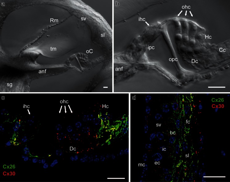
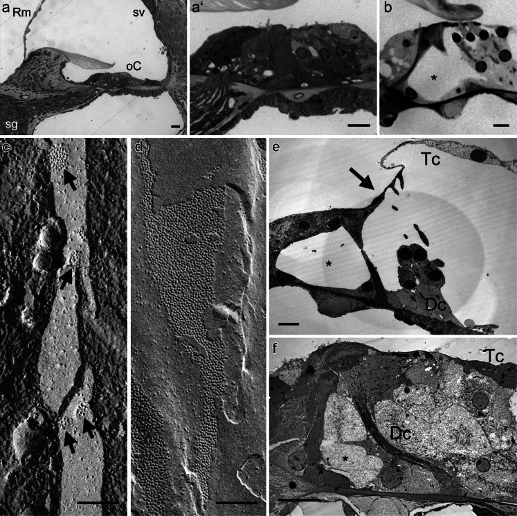
Normal development, function and repair of the sensory epithelia in the inner ear are all dependent on gap junctional intercellular communication. Mutations in the connexin genes GJB2 and GJB6 (encoding CX26 and CX30) result in syndromic and non-syndromic deafness via various mechanisms. Clinical vestibular defects, however, are harder to connect with connexin dysfunction. Cx26 and Cx30 proteins are widely expressed in the epithelial and connective tissues of the cochlea, where they may form homomeric or heteromeric gap junction channels in a cell-specific and spatiotemporally complex fashion. Despite the study of mutant channels and animal models for both recessive and dominant autosomal deafness, it is still unclear why gap junctions are essential for auditory function, and why Cx26 and Cx30 do not compensate for each other in vivo. Cx26 appears to be essential for normal development of the auditory sensory epithelium, but may be dispensable during normal hearing. Cx30 appears to be essential for normal repair following sensory cell loss. The specific modes of intercellular signalling mediated by inner ear gap junction channels remain undetermined, but they are hypothesised to play essential roles in the maintenance of ionic and metabolic homeostasis in the inner ear. Recent studies have highlighted involvement of gap junctions in the transfer of essential second messengers between the non-sensory cells, and have proposed roles for hemichannels in normal hearing. Here, we summarise the current knowledge about the molecular and functional properties of inner ear gap junctions, and about tissue pathologies associated with connexin mutations.
Figures
Similar articles
-
Mice with conditional deletion of Cx26 exhibit no vestibular phenotype despite secondary loss of Cx30 in the vestibular end organs.Hear Res. 2015 Oct;328:102-12. doi: 10.1016/j.heares.2015.07.018. Epub 2015 Jul 29. Hear Res. 2015. PMID: 26232528 Free PMC article.
-
Mutations in the gene for connexin 26 (GJB2) that cause hearing loss have a dominant negative effect on connexin 30.Hum Mol Genet. 2003 Apr 15;12(8):805-12. doi: 10.1093/hmg/ddg076. Hum Mol Genet. 2003. PMID: 12668604
-
Gap junctions in the inner ear: comparison of distribution patterns in different vertebrates and assessement of connexin composition in mammals.J Comp Neurol. 2003 Dec 8;467(2):207-31. doi: 10.1002/cne.10916. J Comp Neurol. 2003. PMID: 14595769
-
Gap junctions and connexins in the inner ear: their roles in homeostasis and deafness.Curr Opin Otolaryngol Head Neck Surg. 2008 Oct;16(5):452-7. doi: 10.1097/MOO.0b013e32830e20b0. Curr Opin Otolaryngol Head Neck Surg. 2008. PMID: 18797288 Review.
-
Recent insights into gap junction biogenesis in the cochlea.Dev Dyn. 2023 Feb;252(2):239-246. doi: 10.1002/dvdy.538. Epub 2022 Sep 26. Dev Dyn. 2023. PMID: 36106826 Review.
Cited by
-
GJB2 and GJB6 gene transcripts in the human cochlea: A study using RNAscope, confocal, and super-resolution structured illumination microscopy.Front Mol Neurosci. 2022 Sep 20;15:973646. doi: 10.3389/fnmol.2022.973646. eCollection 2022. Front Mol Neurosci. 2022. PMID: 36204137 Free PMC article.
-
Mice with conditional deletion of Cx26 exhibit no vestibular phenotype despite secondary loss of Cx30 in the vestibular end organs.Hear Res. 2015 Oct;328:102-12. doi: 10.1016/j.heares.2015.07.018. Epub 2015 Jul 29. Hear Res. 2015. PMID: 26232528 Free PMC article.
-
3D Chromatin Organization Involving MEIS1 Factor in the cis-Regulatory Landscape of GJB2.Int J Mol Sci. 2022 Jun 23;23(13):6964. doi: 10.3390/ijms23136964. Int J Mol Sci. 2022. PMID: 35805969 Free PMC article.
-
Research progress in delineating the pathological mechanisms of GJB2-related hearing loss.Front Cell Neurosci. 2023 Jun 2;17:1208406. doi: 10.3389/fncel.2023.1208406. eCollection 2023. Front Cell Neurosci. 2023. PMID: 37333892 Free PMC article. Review.
-
Purinergic signaling in cochlear supporting cells reduces hair cell excitability by increasing the extracellular space.Elife. 2020 Jan 8;9:e52160. doi: 10.7554/eLife.52160. Elife. 2020. PMID: 31913121 Free PMC article.
References
-
- Ahmad S, Chen S, Sun J, Lin X. Connexins 26 and 30 are co-assembled to form gap junctions in the cochlea of mice. Biochem Biophys Res Commun. 2003;307:362–368. - PubMed
-
- Anselmi F, Hernandez VH, Crispino G, Seydel A, Ortolano S, Roper SD, Kessaris N, Richardson W, Rickheit G, Filippov MA, Monyer H, Mammano F. ATP release through connexin hemichannels and gap junction transfer of second messengers propagate Ca2+ signals across the inner ear. Proc Natl Acad Sci U S A. 2008;105:18770–18775. - PMC - PubMed
-
- Ayad WA, Locke D, Koreen IV, Harris AL. Heteromeric, but not homomeric, connexin channels are selectively permeable to inositol phosphates. J Biol Chem. 2006;281:16727–16739. - PubMed
Publication types
MeSH terms
Substances
Grants and funding
LinkOut - more resources
Full Text Sources
Other Literature Sources
Medical
Molecular Biology Databases
Miscellaneous
