Review
. 2015 Jul;361(1):7-24.
doi: 10.1007/s00441-014-2031-5. Epub 2014 Nov 9.

Inner ear development: building a spiral ganglion and an organ of Corti out of unspecified ectoderm

Affiliations
Review

Inner ear development: building a spiral ganglion and an organ of Corti out of unspecified ectoderm

Bernd Fritzsch et al. Cell Tissue Res. 2015 Jul.

Abstract

The mammalian inner ear develops from a placodal thickening into a complex labyrinth of ducts with five sensory organs specialized to detect position and movement in space. The mammalian ear also develops a spiraled cochlear duct containing the auditory organ, the organ of Corti (OC), specialized to translate sound into hearing. Development of the OC from a uniform sheet of ectoderm requires unparalleled precision in the topological developmental engineering of four different general cell types, namely sensory neurons, hair cells, supporting cells, and general otic epithelium, into a mosaic of ten distinctly recognizable cell types in and around the OC, each with a unique distribution. Moreover, the OC receives unique innervation by ear-derived spiral ganglion afferents and brainstem-derived motor neurons as efferents and requires neural-crest-derived Schwann cells to form myelin and neural-crest-derived cells to induce the stria vascularis. This transformation of a sheet of cells into a complicated interdigitating set of cells necessitates the orchestrated expression of multiple transcription factors that enable the cellular transformation from ectoderm into neurosensory cells forming the spiral ganglion neurons (SGNs), while simultaneously transforming the flat epithelium into a tube, the cochlear duct, housing the OC. In addition to the cellular and conformational changes forming the cochlear duct with the OC, changes in the surrounding periotic mesenchyme form passageways for sound to stimulate the OC. We review molecular developmental data, generated predominantly in mice, in order to integrate the well-described expression changes of transcription factors and their actions, as revealed in mutants, in the formation of SGNs and OC in the correct position and orientation with suitable innervation. Understanding the molecular basis of these developmental changes leading to the formation of the mammalian OC and highlighting the gaps in our knowledge might guide in vivo attempts to regenerate this most complicated cellular mosaic of the mammalian body for the reconstitution of hearing in a rapidly growing population of aging people suffering from hearing loss.


Figures

Fig. 1
Fig. 1
Thin-sheet laser imaging microscopy (TSLIM) and 3D-reconstructions show a developing left ear, viewed from lateral (top row, anterior is to the left) and ventral (bottom row, anterior is to the left). Note the dramatic growth of the cochlea to become the largest duct in the mouse ear exceeding the semicircular canals in length and width. AC, anterior canal crista; C, cochlear duct; HC, horizontal (lateral) canal crista; PC, posterior canal crista; S, saccule; U, utricle. Bar indicates 100 μm. Compiled after (Kopecky et al., 2012).
Fig. 2
Fig. 2
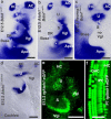
The developing ear has neurosensory precursors that share expression of Bdnf (a-c), whether they develop into hair cells or neurons. In control animals, all sensory epithelia are positive for Bdnf (a), but only the canal cristae and the apex of the cochlea retain Bdnf expression in Atoh1 null mice (b), indicating that the fate of sensory cells is different in different epithelia. In contrast to Atoh1 null, Neurog1 null mice never develop ganglion neurons; show an enlargement of the utricle but a severe reduction of the saccule (c). While only canal cristae and apical precursors remain Bdnf positive in Atoh1 null mice (b), mice homozygotic for Atoh1LacZ show profound expression in sensory precursors nearly equivalent to control mice at this early stage (d). Delaminating neurons from the ear are only positive for Bdnf (asterisks in a and b), but some neurons are also positive for eGFP expression driven by an Atoh1 enhancer element (Vgl in e) that also labels all vestibular hair cells (e). At later stages, this Atoh1-eGFP expression expands not only to all hair cells of OC but also in almost all inner pillar cells (IPC, f). Bar indicates 100 μm (a-e) and 25 μm (f). Compiled after (Fritzsch et al., 2005b, Matei et al., 2005).
Fig. 3
Fig. 3
The normal pattern of OC innervation around birth is shown (a, d) as well as aberrations induced with three mutations (b,c,e). The normal pattern is a very regular set of tunnel crossing fibers (TC) that project to three bundles forming between adjacent Deiter's cells (a,d). Note that fibers as they grow to outer hair cells always turn toward the base (d) and that the majority of radial fibers (RF) end at inner hair cells (IHC). Targeted deletion of Prox1 in SGNs using Nestin-Cre results in aggregation of all tunnel crossing fibers (TC) between the outer pillar cells (OP) and the third row of Deiter's cells (D3). Note that most inner pillar cells (IPC), outer pillar cells (OP) and Deiter's cells remain positive for Prox1, indicating a selective effect of Prox1 expression in SGNs to drive the fiber sorting. Altering the properties of supporting cells in Fgfr3 null mice results in disorganization of afferents to hair cells (c). Most disruptive for the pattern of innervation is the lack of Schwann cells (e). Radial fibers extend from the more centrally migrated SGNs to either bypass entirely the hair cells of the OC to end up at the lateral wall (LW) or to form a disorganized innervation of the OC. Bar indicates 20 μm (a-c) and 100 μm (d,e). Data are from (Mao et al., 2014, Yang et al., 2011).
Fig. 4
Fig. 4
Spiral ganglion axons reach the cochlear nuclei around E12.5 (a) and are from the initial projection clearly separated from vestibular axons of the posterior vertical canal crista (PC). Vestibular afferents extend early past the boundaries of the cochlear nuclei into the brainstem and the cerebellum whereas cochlear afferents in the anteroventral (AVCN) and dorsal cochlear nuclei (DCN). A segregated projection of base and apex is already established in E14.5 embryos (b) but appears to become refined in early neonates (c). Abbreviations: AVCN, anteroventral cochlear nucleus; Cne, cochlear nerve; Co, cochlear application; DCN, dorsal cochlear nucleus; eff, olivocochlear efferent bundle; PC, posterior canal crista application. Bar indicates 100 μm. Modified after (Fritzsch et al., 2005a, Jahan et al., 2010a, Maklad and Fritzsch, 2003).
Fig. 5
Fig. 5
The organ of Corti (OC) is often depicted by most theoretical papers as a simple checkerboard of alternating hair cells and supporting cells (a), in which each hair cell is surrounded by four supporting cells and each supporting cell is surrounded by four hair cells. This checkerboard pattern exists, however, only in the ‘outer compartment’ of the OC with alternating rows of outer hair cells (OHC) and Deiter's cells/outer pillar cells (D1-3; OPC). This outer compartment is separated by a single row of adjacent inner pillar cells (IPC) from the inner compartment, consisting of two alternating cell types, the inner hair cell (IHC) and the inner phalangeal cells (IPhC). Note that inner phalangeal cells seem to be organized in pairs flanking either medially or laterally the IHCs according to more recent TEM data. Note also the numerical match of outer compartment elements in humans whereas neither IHC nor IPC fit to the numbers of the outer compartments (b). In particular the continuity of IPCs in a single row and the fact that two supporting cells (IPC, OPC) are adjacent to and touching each other is difficult to reconcile with simple interpretations of the Delta/Notch interaction of lateral inhibition. Modified after (Slepecky, 1996, Spoendlin and Schrott, 1988, Zetes et al., 2012).
Fig. 6
Fig. 6
The distribution of OC cells and the origin and hypothetical gradients of several diffusible factors (Fgfs, BMP4) are shown in control (a,b) and Neurod1 null ears (c,d). Note that the conversion of outer hair cells into Fgf8 positive hair cells in Neurod1 null mice results in transformation of surrounding Deiter's cells into pillar cell like cells (white arrows in d), indicating that a second center of Fgf8 diffusion can disrupt the cellular mosaic of the outer compartment of the OC (c,d). In essence, diffusible factors cooperate with lateral inhibition and selected expression of other genes to coordinate the normal development of the OC. Misexpression of only one diffusible factor (Fgf8 in some OHCs) can override all other interactions leading to a disrupted development of the OC (Jahan et al., 2010b). Bar in b and d indicates 100 μm. a,c,d, is modified after (Groves and Fekete, 2012, Jahan et al., 2013), b is unpublished data.

References

    1. Ahmed M, Xu J, Xu PX. EYA1 and SIX1 drive the neuronal developmental program in cooperation with the SWI/SNF chromatin-remodeling complex and SOX2 in the mammalian inner ear. Development. 2012;139:1965–1977. - PMC - PubMed
    1. Alam SA, Robinson BK, Huang J, Green SH. Prosurvival and proapoptotic intracellular signaling in rat spiral ganglion neurons in vivo after the loss of hair cells. The Journal of comparative neurology. 2007;503:832–852. - PubMed
    1. Appler JM, Goodrich LV. Connecting the ear to the brain: Molecular mechanisms of auditory circuit assembly. Prog Neurobiol. 2011;93:488–508. - PMC - PubMed
    1. Appler JM, Lu CC, Druckenbrod NR, Yu WM, Koundakjian EJ, Goodrich LV. Gata3 is a critical regulator of cochlear wiring. The Journal of neuroscience : the official journal of the Society for Neuroscience. 2013;33:3679–3691. - PMC - PubMed
    1. Basch ML, Ohyama T, Segil N, Groves AK. Canonical Notch signaling is not necessary for prosensory induction in the mouse cochlea: insights from a conditional mutant of RBPjkappa. J Neurosci. 2011;31:8046–8058. - PMC - PubMed

Publication types

LinkOut - more resources