

doi: 10.1002/cbic.201402512.
Epub 2014 Nov 7.
Identification and characterization of a small-molecule inhibitor of death-associated protein kinase 1
Affiliations
- PMID: 25382253
- PMCID: PMC4431585
- DOI: 10.1002/cbic.201402512
Item in Clipboard
Identification and characterization of a small-molecule inhibitor of death-associated protein kinase 1
Chembiochem.
.
No abstract available
Keywords: drug discovery; enzymes; inhibitors; phosphorylation; protein structures.
Figures
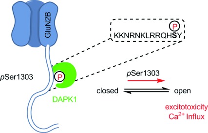
Proposed mechanism for GluN2Bs increased excitotoxicity after phosphorylation by DAPK1. After phosphorylation of Ser1303 DAPK1 remains bound to the phosphorylation site with its catalytic domain, thereby mediating increased calcium influx.
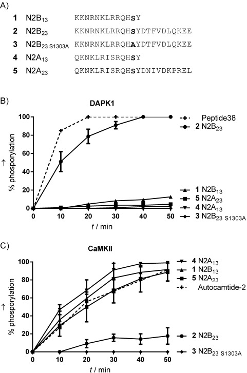
Comparison of peptide substrates containing Ser1303 (GluN2B) or Ser1291 (GluN2A). A) Alignment of peptides used in in vitro phosphorylation studies. B) Phosphorylation by DAPK1. C) Phosphorylation by CaMKII. Control substrates are shown with dashed lines. Degrees of phosphorylation were assessed by analytical HPLC (n=3, mean±SD). Concentration: peptide 50 μm , ATP 100 μm, kinase concentration was adjusted to fit the timescale. For further details see the Supporting Information.
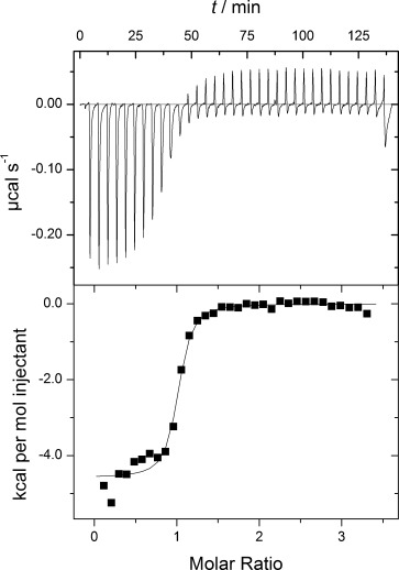
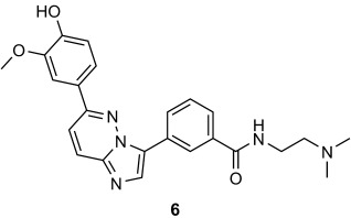
Representative ITC of the catalytic domain of DAPK1 (residues 1–285) into 6. ITC reveals an equilibrium dissociation constant (Kd) of 0.24± 0.09 μm (n=4).
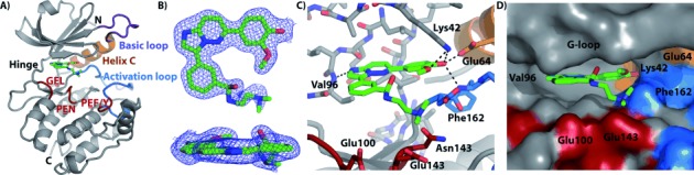
DAPK1–CPR005231 (6) co-crystal structure (PDB ID: 4TXC) A) Structural model and ligand binding site showing the basic loop (purple), helix C (orange), activation loop (blue), substrate binding motif residues (red), and ligand (green). B) Electron density map contoured at 1 σ for the ligand shown from two perspectives. (2Fo−Fc) C) Detail of the ligand-binding site showing key interactions and proximity to DAPK1 substrate-binding motif residues. D) Surface-filled representation of the ligand-binding site illustrating nearby pockets for potential ligand modification.
References
Publication types
MeSH terms
Substances
Grants and funding
LinkOut - more resources
Full Text Sources
Other Literature Sources
