Review
. 2015 Feb 15;24(4):405-16.
doi: 10.1089/scd.2014.0442. Epub 2014 Dec 18.

Cancer stem cell division: when the rules of asymmetry are broken

Affiliations
Review

Cancer stem cell division: when the rules of asymmetry are broken

Subhas Mukherjee et al. Stem Cells Dev. .

Abstract

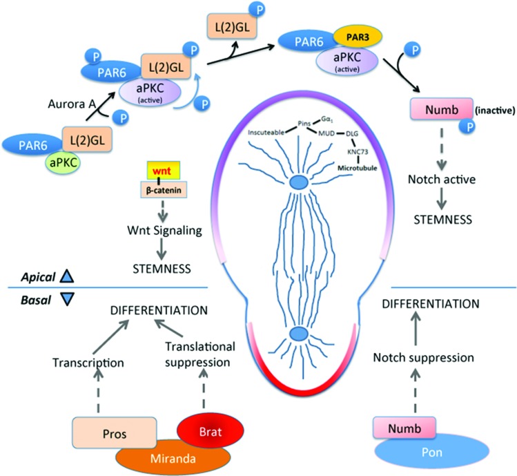
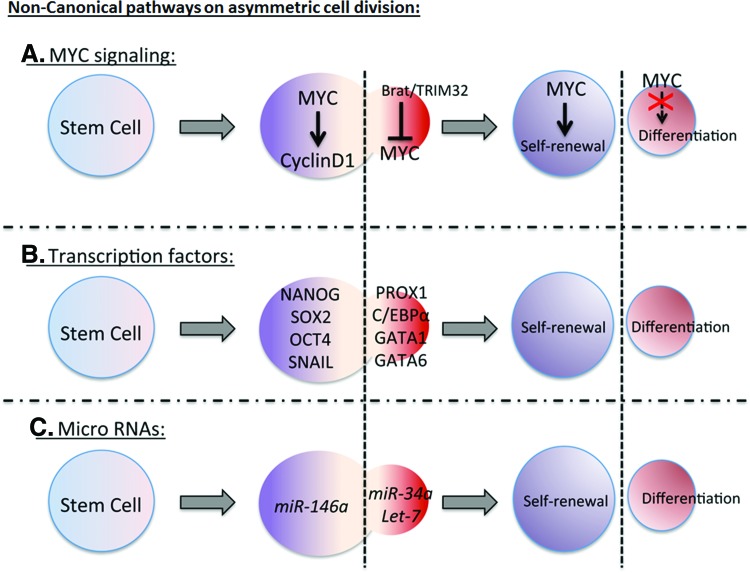
Asymmetric division of stem cells is a highly conserved and tightly regulated process by which a single stem cell produces two daughter cells and simultaneously directs the differential fate of both: one retains its stem cell identity while the other becomes specialized and loses stem cell properties. Coordinating these events requires control over numerous intra- and extracellular biological processes and signaling networks. In the initial stages, critical events include the compartmentalization of fate determining proteins within the mother cell and their subsequent passage to the appropriate daughter cell. Disturbance of these events results in an altered dynamic of self-renewing and differentiation within the cell population, which is highly relevant to the growth and progression of cancer. Other critical events include proper asymmetric spindle assembly, extrinsic regulation through micro-environmental cues, and noncanonical signaling networks that impact cell division and fate determination. In this review, we discuss mechanisms that maintain the delicate balance of asymmetric cell division in normal tissues and describe the current understanding how some of these mechanisms are deregulated in cancer. The universe is asymmetric and I am persuaded that life, as it is known to us, is a direct result of the asymmetry of the universe or of its indirect consequences. The universe is asymmetric. -Louis Pasteur.


Figures

<b>FIG. 1.</b>
FIG. 1.
During asymmetric cell division, two distinct molecular programs take place on the apical and basal pole. Apical pole: At the apical side, aPKC/PAR6/PAR3 complex formation initiates during interphase, giving apical pole the identity of self-renewal. Aurora A protein kinase phosphorylates aPKC leading to its activation. Active aPKC phosphorylates L(2)GL which releases L(2)GL from the complex and PAR3 enters. This complex phosphorylates Numb releasing it from the apical membrane and increasing Numb concentration at the basal side. This keeps the Notch signal active on the apical side causing stemness. Wnt signal also takes part in the self-renewal, though the detailed mechanism is not known. In metaphase and telophase, apical microtubule arrangement is maintained by Inscuteable/Pins/Gαi complex. Cytoskeletal adapter protein binds with Pins and Gαi whereas Mud forms a complex with Dlg and Knc73. These two complexes come together that arranges the microtubules attached to Knc73. Basal pole: Adapter protein Miranda on the basal pole binds with Prospero and Brat. Degradation of Miranda releases transcription factor Prospero that turns on the genes driving differentiation. Brat on the other hand, acts as a translational repressor, possibly suppressing protein production needed for proliferation. brat ortholog TRIM32 also transports cMYC to endosomes for its degradation. Higher accumulation of NUMB on the basal membrane leads to the degradation of NOTCH1 inside endosomes. This suppresses Notch signal and its proliferative effect on the basal side. aPKC, atypical protein kinase C. Color images available online at www.liebertpub.com/scd
<b>FIG. 2.</b>
FIG. 2.
Noncanonical signaling pathways regulate asymmetric cell division. (A) Various growth-signaling pathways affect the asymmetric cell division balance. Progrowth regulators such as MYC shift the asymmetric cell division balance toward self-renewal, through activating CYCLIN D1. On the basal side, Drosophila Brat or mammalian TRIM32 attenuates MYC function to start differentiation signal. (B) Dysregulation of pluripotency and self-renewal transcription factors such as NANOG, SOX2, OCT4, and SNAIL also initiate symmetric stem cell proliferation. Transcription factors such as PROX1, C/EBP alpha, GATA1, GATA6 are expressed in the differentiated daughter cell. (C) MicroRNAs also regulate asymmetric cell division by affecting stem cell renewal and differentiation. miR-146a drives stem cell renewal, whereas miR-34a and Let-7 drive differentiation. Color images available online at www.liebertpub.com/scd

References

    1. Knoblich JA. (2010). Asymmetric cell division: recent developments and their implications for tumour biology. Nat Rev Mol Cell Biol 11:849–860 - PMC - PubMed
    1. Yong KJ. and Yan B. (2011). The relevance of symmetric and asymmetric cell divisions to human central nervous system diseases. J Clin Neurosci 18:458–463 - PubMed
    1. Conklin EG. (1905). The mutation theory from the standpoint of cytology. Science 21:525–529 - PubMed
    1. Yamashita YM, Mahowald AP, Perlin JR. and Fuller MT. (2007). Asymmetric inheritance of mother versus daughter centrosome in stem cell division. Science 315:518–521 - PMC - PubMed
    1. Bultje RS, Castaneda-Castellanos DR, Jan LY, Jan YN, Kriegstein AR. and Shi SH. (2009). Mammalian Par3 regulates progenitor cell asymmetric division via notch signaling in the developing neocortex. Neuron 63:189–202 - PMC - PubMed

Publication types

LinkOut - more resources