. 2015 Feb;36(2):210-21.
doi: 10.1002/humu.22725.

Functional classification of BRCA2 DNA variants by splicing assays in a large minigene with 9 exons

Affiliations

Functional classification of BRCA2 DNA variants by splicing assays in a large minigene with 9 exons

Alberto Acedo et al. Hum Mutat. 2015 Feb.

Abstract

Numerous pathogenic DNA variants impair the splicing mechanism in human genetic diseases. Minigenes are optimal approaches to test variants under the splicing viewpoint without the need of patient samples. We aimed to design a robust minigene construct of the breast cancer gene BRCA2 in order to investigate the impact of variants on splicing. BRCA2 exons 19-27 (MGBR2_ex19-27) were cloned in the new vector pSAD. It produced a large transcript of the expected size (2,174 nucleotides) and exon structure (V1-ex19-27-V2). Splicing assays showed that 18 (17 splice-site and 1 silencer variants) out of 40 candidate DNA variants induced aberrant patterns. Twenty-four anomalous transcripts were accurately detected by fluorescent-RT-PCR that were generated by exon-skipping, alternative site usage, and intron-retention events. Fourteen variants induced major anomalies and were predicted to disrupt protein function so they could be classified as pathogenic. Furthermore, minigene mimicked previously reported patient RNA outcomes of seven variants supporting the reproducibility of minigene assays. Therefore, a relevant fraction of variants are involved in breast cancer through splicing alterations. MGBR2_ex19-27 is the largest reported BRCA2 minigene and constitutes a valuable tool for the functional and clinical classification of sequence variations.

Keywords: BRCA2; hereditary breast and ovarian cancer; minigenes; splicing; unclassified variants.


Figures

Figure 1
Figure 1
Structures and functional analysis of the splicing vector pSAD and the minigene MGBR2_ex19–27. A: The pSAD vector contains a SV40 transcription promoter, two constituve exons (V1 and V2), and two selection markers: ampicillin resistance and β-galactosidase (LacZ) with a multiple cloning site (mcs). Specific primers to amplify minigene transcripts are indicated by arrows in V1 and V2 exons. B: Structure of the minigene MGBR2_ex19–27: [IVS18 (247 bp) - EX19 (156 bp) - IVS19 (398 bp) - EX20 (145 bp) - IVS20 (207 bp) // IVS20 (90 bp) - EX21 (122 bp) - IVS21 (262 bp) // IVS21 (262 bp) - EX22 (199 bp) - IVS22 (234 bp) - EX23 (164 bp) - IVS23 (93 bp) - EX24 (139 bp) - IVS24 (147 bp)// IVS24 (271 bp) - EX25 (245 bp) - IVS25 (431 bp) // IVS25 (341 bp) - EX26 (147 bp) - IVS26 (344 bp) // IVS26 (221 bp) - EX27 (723 bp)]. The expected splicing reactions in eukaryotic cells are indicated by arrows. The acceptor site and part of exon V2 was replaced by that with 221 bp of intron 26 and 723 bp of exon 27 with its corresponding acceptor site but maintaining the V2 sequence to anneal specific reverse RT-PCR primer. C: Splicing Functional Assays of the pSAD® v5.0 vector, the intermediate minigenes MGBR2i_EX19–24, MG BR2i_EX19–25, MG BR2i_EX19–26 and the final construct MGBR2_EX19–27. RNA was retrotranscribed and amplified with vector specific primers. Sizes of each transcript are indicated below each band.
Figure 2
Figure 2
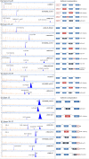
Capillary electrophoresis of fluorescent RT-PCR products from wild-type MGBR2_ex19–27 and derived mutant minigenes. RT-PCR products were amplified with one FAM-primer forward (priming on exon V1) or reverse (V2) of the pSAD vector and one exonic BRCA2 primer. All samples were run on an ABI3130 sequencer with Genescan LIZ1200 (“orange/faint”peaks) as size standard. Screenshots of electropherograms visualized with the Peak Scanner software are shown. Fragment sizes and relative fluorescent units are indicated on the x- and y-axes, respectively. wt designates the expected canonical transcript with each primer pair. A: Analysis of exons 19 and 20 (primers RTpSAD-FW and RTBR2_ex22-RV); B: exons 21 and 22 (RTpSAD-FW and RTBR2_ex23-RV); C: exons 23 and 24 (RTpSAD-FW and RTBR2_ex25-RV); D: exon 25 (RTBR2_ex24-FW and RTpSAD-RV); E: exons 26 and 27 (RTBR2_ex25-FW and RTpSAD-RV). The exon composition and the splicing reaction of the different isoforms are shown on the right. Transcripts that were not fully characterized (ins23 or 436-nt) are not represented.
Figure 3
Figure 3
Effect of Nonsense-mediated decay inhibitors on splicing outcome of DNA variant c.8948_8953+5del. Wild type and mutant minigenes were transfected into HeLa cells and treated (+CHX) or not (-CHX) with cycloheximide. RNA was retrotranscribed and cDNA was amplified with primers RTPSAD-FW and RTBR2_ex23-RV. A: An agarose gel electrophoresis is shown on the left where the expected canonical transcript is indicated by an arrow and asterisks indicate novel transcripts or significant changes in transcript amounts after NMD inhibition. Electropherograms of fluorescent RT-PCR products of variant c.8948_8953+5del are shown on the right. B: Effect of NMD on the relative proportions of each transcript with (grey bars) and without cycloheximide (white bars). Values are averages of three independent experiments. The size of each transcript is indicated between parentheses.
Figure 4
Figure 4
Mapping of splicing regulatory sequences by functional assays of exonic microdeletions of exons 19 (A) and 20 (B). Deletions are designated according to the HGVS nomenclature. Arrows indicate possible abnormal transcripts.

References

    1. Acedo A, Sanz DJ, Duran M, Infante M, Perez-Cabornero L, Miner C, Velasco EA. Comprehensive splicing functional analysis of DNA variants of the BRCA2 gene by hybrid minigenes. Breast Cancer Res. 2012;14:R87. - PMC - PubMed
    1. Baralle D, Lucassen A, Buratti E. Missed threads. The impact of pre-mRNA splicing defects on clinical practice. EMBO Rep. 2009;10:810–816. - PMC - PubMed
    1. Baralle M, Skoko N, Knezevich A, De CL, Motti D, Bhuvanagiri M, Baralle D, Buratti E, Baralle FE. NF1 mRNA biogenesis: effect of the genomic milieu in splicing regulation of the NF1 exon 37 region. FEBS Lett. 2006;580:4449–4456. - PubMed
    1. Bonatti F, Pepe C, Tancredi M, Lombardi G, Aretini P, Sensi E, Falaschi E, Cipollini G, Bevilacqua G, Caligo MA. RNA-based analysis of BRCA1 and BRCA2 gene alterations. Cancer Genet Cytogenet. 2006;170:93–101. - PubMed
    1. Bonnet C, Krieger S, Vezain M, Rousselin A, Tournier I, Martins A, Berthet P, Chevrier A, Dugast C, Layet V, Rossi A, Lidereau R, et al. Screening BRCA1 and BRCA2 unclassified variants for splicing mutations using reverse transcription PCR on patient RNA and an ex vivo assay based on a splicing reporter minigene. J Med Genet. 2008;45:438–446. - PubMed

Publication types