. 2012:159:357-370.
doi: 10.1039/C2FD20062G.

The role of the amorphous phase on the biomimetic mineralization of collagen

Affiliations

The role of the amorphous phase on the biomimetic mineralization of collagen

Fabio Nudelman et al. Faraday Discuss. 2012.

Abstract

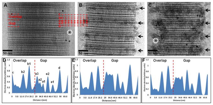
Bone is a hierarchically structured composite material whose basic building block is the mineralized collagen fibril, where the collagen is the scaffold into which the hydroxyapatite (HA) crystals nucleate and grow. Understanding the mechanisms of hydroxyapatite formation inside the collagen is key to unravelling osteogenesis. In this work, we employed a biomimetic in vitro mineralization system to investigate the role of the amorphous precursor calcium phosphate phase in the mineralization of collagen. We observed that the rate of collagen mineralization is highly dependent on the concentration of polyaspartic acid, an inhibitor of hydroxyapatite nucleation and inducer of intrafibrillar mineralization. The lower the concentration of the polymer, the faster the mineralization and crystallization. Addition of the non-collagenous protein C-DMP1, a nucleator of hydroxyapatite, substantially accelerates mineral infiltration as well as HA nucleation. We have also demonstrated that Cu ions interfere with the mineralization process first by inhibiting the entry of the calcium phosphate into the collagen, and secondly by stabilizing the ACP, such that it does not convert into HA. Interestingly, under these conditions mineralization happens preferentially in the overlap regions of the collagen fibril. Our results show that the interactions between the amorphous precursor phase and the collagen fibril play an important role in the control over mineralization.


Figures

Fig. 1
Fig. 1
CryoTEM images of collagen mineralized with calcium phosphate for 24 h in presence of different concentrations of pAsp, on cryoTEM grids made of Au. A. 1.5 μg/ml of pAsp. White arrows: apatite crystals nucleating and growing within the amorphous calcium phosphate phase. B. 3 μg/ml of pAsp. C. 6 μg/ml of pAsp. D. 10 μg/ml of pAsp. White arrows: amorphous calcium phosphate particles infiltrating into the collagen. Panel D was adapted from Nudelman, F. et al., Nature Mater., 2010, 9, 1004–1009. Reprinted with permission from Macmillan Publishers Ltd. Nature Materials, www.nature.com/nmat, Copyright 2010.
Fig. 2
Fig. 2
CryoTEM images of collagen at different stages of mineralization in presence of 10 μg/ml of pAsp, on cryoTEM grids made of Au. A. Mineralization for 24 h. B. Mineralization for 48 h. C. Mineralization for 72 h. D. Collagen fibril containing two regions, one well organized (black circle area) and one poorly organized (black circle area). The well organized area is partially mineralized with amorphous calcium phosphate and apatite, while the poorly organized one contains only few, randomly oriented apatite crystals (white dashed circle). Dashed black circle: 10 nm gold marker. E. A partially mineralized collagen fibril, where deformation caused by the presence of the mineral can be observed. Dashed-lines: Region with least (dashed-line 1) and most amount of mineral (dashed-line 2) from where the cross-sectional area of the fibril was calculated. Panel E was adapted from Nudelman, F. et al., Nature Mater., 2010, 9, 1004–1009. Reprinted with permission from Macmillan Publishers Ltd. Nature Materials, www.nature.com/nmat, Copyright 2010.
Fig. 3
Fig. 3
CryoTEM images of collagen at different stages of mineralization in presence of 10 μg/ml of pAsp, on cryoTEM grids made of Cu. A. Mineralization for 24 h. White circle: amorphous calcium phosphate particles associated to the collagen. B. Mineralization for 48 h. White circle: amorphous calcium phosphate particles associated to the overlap region of collagen. Black circle: 10 nm gold markers. C. Mineralization for 72 h. White circle: amorphous calcium phosphate particles associated to the overlap region of collagen. D. Low-dose selected-area electron diffraction of C, showing that the mineral is still amorphous. E. Cryo-energy dispersive X-ray spectroscopy (cryoEDX) measurement of C, confirming that the precipitates are indeed composed of calcium phosphate.
Fig. 4
Fig. 4
Cryo-electron tomography of a collagen fibril mineralized for 48 h in the presence of 10 μg/ml of pAsp, on a Cu grid. A. Two-dimensional cryoTEM image. B. Slice from a section of the reconstructed 3-dimensional volume, showing calcium phosphate particles inside the collagen (white circles). C. Computer-generated 3-dimensional visualization of the reconstruction, where the collagen fibril is depicted in white and the calcium phosphate precipitates in red. Inset: only half of the collagen fibril is shown, revealing the calcium phosphate precipitates inside the fibril, predominantly in the overlap region. D. Graph showing the distribution of calcium phosphate within a 67 nm repeat.
Fig. 5
Fig. 5
Uranyl acetate map of collagen during the early stages of mineralization. A. Non-mineralized collagen. White circle: 10 nm gold marker. B. Collagen mineralized for 24 h in presence of 10 μg/ml of pAsp, on Au grids. C. Collagen mineralized for 24 h in presence of 10 μg/ml of pAsp, on Cu grids. White circle: 10 nm gold marker. D. Intensity profile of A, non-mineralized collagen. Dashed line: border between gap and overlap regions (C-terminus). E. Intensity profile of B, collagen mineralized for 24 h on Au grids. Dashed line: border between gap and overlap regions (C-terminus). F. Intensity profile of C, collagen mineralized for 24 h on Cu grids. Dashed line: border between gap and overlap regions (C-terminus). Panels A and D were adapted from Nudelman, F. et al., Nature Mater., 2010, 9, 1004–1009. Reprinted with permission from Macmillan Publishers Ltd. Nature Materials, www.nature.com/nmat, Copyright 2010.
Fig. 6
Fig. 6
A. CryoTEM image of collagen mineralized for 4 weeks in presence of 10 μg/ml of pAsp on Cu grids. The calcium phosphate is still amorphous, as shown by the LDSAED (inset). B. TEM image of dried collagen mineralized for 4 weeks in presence of 10 μg/ml of pAsp on Cu grids. Even after freeze-drying, the mineral shows no signs if crystallinity, as shown by the LDSAED (inset). C. TEM image of dried collagen mineralized for 2 weeks without additives, on a Cu grid. The surface of the collagen and of the carbon support film on the grid are completely covered with apatite crystals. Inset: LDSAED, showing the (002) and (211) reflections of apatite. D. TEM image of dried HA crystals precipitated in presence of 10 μg/ml of pAsp on a Cu grid after 2 weeks of reaction, without collagen. Inset: LDSAED, showing the (002) and (211) reflections of apatite.
Fig. 7
Fig. 7
CryoTEM images of collagen mineralized in presence of 15 μg/ml of C-DMP1 on Au and Cu grids. A. Collagen mineralized for 24 h in presence of 15 μg/ml of C-DMP1 on Au grids. Black circle: 10 nm gold markers. B. Collagen mineralized for 24 h in presence of 15 μg/ml of C-DMP1 and 10 μg/ml of pAsp on Au grids. Black circle: 10 nm gold markers. C. Collagen mineralized for 24 h in presence of 15 μg/ml of C-DMP1 on Cu grids. Black arrows: globular structures of calcium phosphate on the surface of collagen. Black circle: 10 nm gold markers. D. Collagen mineralized for 24 h in presence of 15 μg/ml of C-DMP1 and 10 μg/ml of pAsp on Cu grids. Black circle: 10 nm gold markers.

References

    1. Weiner S, Wagner HD. Annu Rev Mater Sci. 1998;28:271–298.
    1. Glimcher MJ, Muir H. Philosophical Transactions of the Royal Society of London Series B, Biological Sciences. 1984;304:479–508. - PubMed
    1. Hodge AJ, Petruska JA. In: Aspects of Protein Structure. Ramachandran GN, editor. Academic Press; New York: 1963. pp. 289–300.
    1. Miller A. Philosophical Transactions of the Royal Society of London Series B, Biological Sciences. 1984;304:455–477. - PubMed
    1. Orgel JPRO, Irving TC, Miller A, Wess TJ. Proceedings of the National Academy of Sciences of the United States of America. 2006;103:9001–9005. - PMC - PubMed