. 2014 Oct 29;14(1):96.
doi: 10.1186/s12935-014-0096-6. eCollection 2014.

Ursolic acid induces cell cycle arrest and apoptosis of gallbladder carcinoma cells

Affiliations

Ursolic acid induces cell cycle arrest and apoptosis of gallbladder carcinoma cells

Hao Weng et al. Cancer Cell Int. .

Abstract

Background: Ursolic acid (UA), a plant extract used in traditional Chinese medicine, exhibits potential anticancer effects in various human cancer cell lines in vitro. In the present study, we evaluated the anti-tumoral properties of UA against gallbladder carcinoma and investigated the potential mechanisms responsible for its effects on proliferation, cell cycle arrest and apoptosis in vitro.

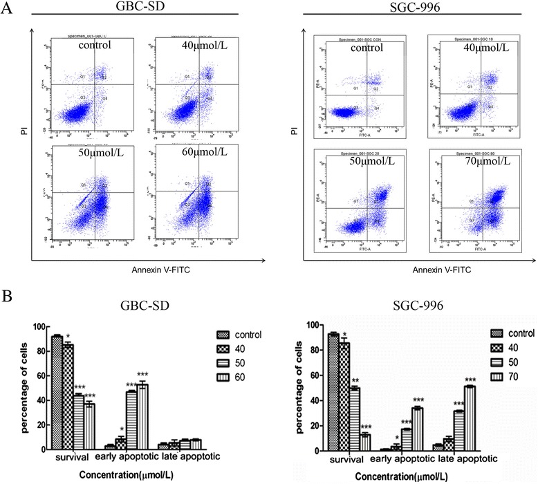
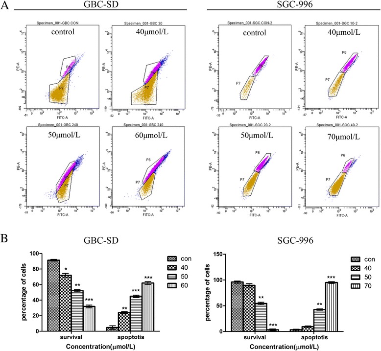
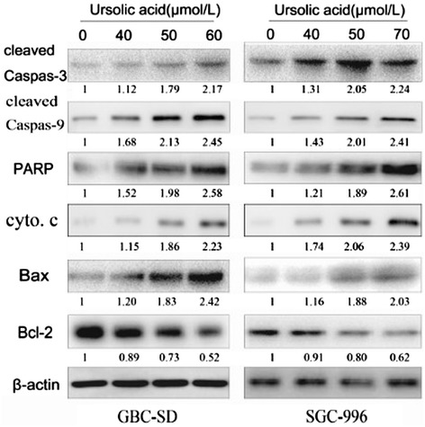
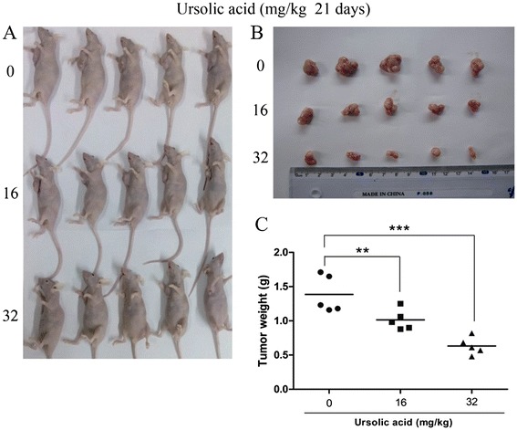
Methods: The anti-tumor activity of UA against GBC-SD and SGC-996 cells was assessed using MTT and colony formation assays. An annexin V/PI double-staining assay was used to detect cell apoptosis. Cell cycle changes were detected using flow cytometry. Rhodamine 123 staining was used to assess the mitochondrial membrane potential (ΔΨm) and validate UA's ability to induce apoptosis in both cell lines. The effectiveness of UA in gallbladder cancer was further verified in vivo by establishing a xenograft GBC model in nude mice. Finally, the expression levels of cell cycle- and apoptosis-related proteins were analyzed by western blotting.

Results: Our results suggest that UA can significantly inhibit the growth of gallbladder cancer cells. MTT and colony formation assays indicated dose-dependent decreases in cell proliferation. S-phase arrest was observed in both cell lines after treatment with UA. Annexin V/PI staining suggested that UA induced both early and late phases of apoptosis. UA also decreased ΔΨm and altered the expression of molecules regulating the cell cycle and apoptosis. In vivo study showed intraperitoneally injection of UA can significantly inhibited the growth of xenograft tumor in nude mice and the inhibition efficiency is dose related. Activation of caspase-3,-9 and PARP indicated that mitochondrial pathways may be involved in UA-induced apoptosis.

Conclusions: Taken together, these results suggest that UA exhibits significant anti-tumor effects by suppressing cell proliferation, promoting apoptosis and inducing 7cell cycle arrest both in vitro and in vivo. It may be a potential agent for treating gallbladder cancer.

Keywords: Apoptosis; Cell cycle; Gallbladder cancer; Mitochondrial-mediated pathway; Proliferation; Ursolic acid.


Figures

Figure 1
Figure 1
UA inhibits proliferation in GBC cells. (A) The chemical structure of UA. (B) GBC-SD and SGC-996 cells were treated with various concentrations of UA for 24, 48 or 72 h. Effects on cell proliferation were determined using a MTT assay. Each value represents the mean ± SD (n = 3). (C-D) UA inhibits colony formation in GBC cells. GBC-SD and SGC-996 cells were treated with different doses of UA (8, 16 or 32 μmol/L) and were allowed to form colonies in fresh medium for 14 days. The photomicrographic differences and number of colonies (mean ± SD, n = 3) in colony formation are shown. Significant differences from the control are indicated by *p < 0.05 and **p < 0.01.
Figure 2
Figure 2
UA induces cell cycle arrest at the S phase in GBC cells. GBC-SD and SGC-996 cells were treated with different concentrations of UA for 48 h. (A) The cell cycle distribution of treated cells was determined using flow cytometry. (B) The data are expressed as the mean ± SD (n = 3), with results representative of 3 independent experiments shown. *p < 0.05, **p < 0.01 and ***p < 0.001 vs. the control group.
Figure 3
Figure 3
UA induces apoptosis in GBC cells. GBC-SD and SGC-996 cells were treated with different concentrations of UA for 48 h. (A) Flow cytometric analysis of UA-induced apoptosis in GBC cells using annexin V-FITC/PI staining. Cells in the lower right quadrant represent early apoptotic cells, and those in the upper right quadrant represent late apoptotic cells. (B) The percentage of apoptotic cells is presented as the mean ± SD. The data are representative of 3 similar experiments. Significant differences from the control are indicated by *p < 0.05, **p < 0.01 and ***p < 0.001.
Figure 4
Figure 4
UA affects ΔΨ m in GBC cells. GBC-SD and SGC-996 cells were treated with UA for 48 h and then stained with the membrane-sensitive probe Rhodamine 123. (A) Rhodamine retention was measured by flow cytometry. The results are representative of 3 independent experiments. (B) The corresponding linear diagram shows the percentages of Rhodamine 123-negative cells as the mean ± SD (n = 3). *p < 0.05, **p < 0.01 and ***p < 0.001 vs. the control group.
Figure 5
Figure 5
UA modulates the expression of cell cycle- and apoptosis-related proteins in GBC cells. Western blot analysis of protein extracts from GBC-SD and SGC-996 cells treated with different doses of UA for 48 h. The expression levels of cleaved caspase-3, caspase-9, PARP, cyto c, Bax and Bcl-2 were analyzed. β-Actin was used as a loading control. The results are representative of 3 independent experiments.
Figure 6
Figure 6
UA suppressed the growth of tumor in nude mice injected with GBC-SD cells. (A) GBC-SD cells were subcutaneously injected into the right flank of the nude mice; The mice were then administered 0.2 mL of vehicle (10% DMSO and 90% PBS) or UA (16 mg/kg and 32 mg/kg) intraperitoneally everyday for up to 22 days. Photos of 5 representative mice (n = 10) from each group were presented to show the sizes of the resulting tumors; (B,C) Tumors were excised from the animals and weighed. *P < 0.05 **P < 0.01 vs. the control group.

References

    1. Miller G, Jarnagin WR. Gallbladder carcinoma. Eur J Surg Oncol. 2008;34(3):306–312. doi: 10.1016/j.ejso.2007.07.206. - DOI - PubMed
    1. Tan Z, Li M, Wu W, Zhang L, Ding Q, Wu X, Mu J, Liu Y. NLK is a key regulator of proliferation and migration in gallbladder carcinoma cells. Mol Cell Biochem. 2012;369(1–2):27–33. doi: 10.1007/s11010-012-1365-0. - DOI - PubMed
    1. Rifatbegovic Z, Mesic D, Ljuca F, Zildzic M, Morankic M. Incidence and surgical treatment of cancer in gallbladder. Med Arh. 2007;61(1):30–33. - PubMed
    1. Wang J, Liu L, Qiu H, Zhang X, Guo W, Chen W, Tian Y, Fu L, Shi D, Cheng J, Huang W, Deng W. Ursolic acid simultaneously targets multiple signaling pathways to suppress proliferation and induce apoptosis in colon cancer cells. PLoS ONE. 2013;8(5):e63872. doi: 10.1371/journal.pone.0063872. - DOI - PMC - PubMed
    1. Wang JS, Ren TN, Xi T. Ursolic acid induces apoptosis by suppressing the expression of FoxM1 in MCF-7 human breast cancer cells. Med Oncol. 2012;29(1):10–15. doi: 10.1007/s12032-010-9777-8. - DOI - PubMed