

doi: 10.1016/j.mmcr.2014.09.004.
eCollection 2014 Oct.
Successful therapy of progressive rhino-orbital mucormycosis caused by Rhizopus arrhizus with combined and sequential antifungal therapy, surgery and hyperbaric therapy
Affiliations
- PMID: 25383316
- PMCID: PMC4223823
- DOI: 10.1016/j.mmcr.2014.09.004
Item in Clipboard
Successful therapy of progressive rhino-orbital mucormycosis caused by Rhizopus arrhizus with combined and sequential antifungal therapy, surgery and hyperbaric therapy
Med Mycol Case Rep.
.
Abstract
We present a case of rhino-orbitary mucormycosis which progressed despite liposomal amphotericin and early surgical debridement. Combined echinocandin and high dose liposomal amphotericin, repeated debridement, prolonged therapy with hyperbaric oxygen and continued therapy with posaconazole, along with strict diabetic control, allowed cure without disfigurement.
Keywords: Amphotericin B; Diabetes mellitus; Hyperbaric oxygen; Posazonale; Rhizopus arrhizu; Zygomycosis.
Figures
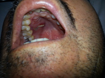
Hard palate befote treatment. Linear ulceration with necrotic mucosa.
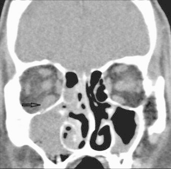
Pretreatment coronal CT: right maxillary sinus opacification with intraorbital extension without bone erosion. Arrow: thickening of inferior rectus muscle and infiltration of adjacent fat.
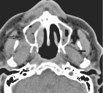
Axial CT: extension of infection in the buccal space and masticator space. Hollow arrow: hyperattenuation right retromaxillary fat pad. Small arrow: thickening masticator space musculature and obliteration of the fat planes.
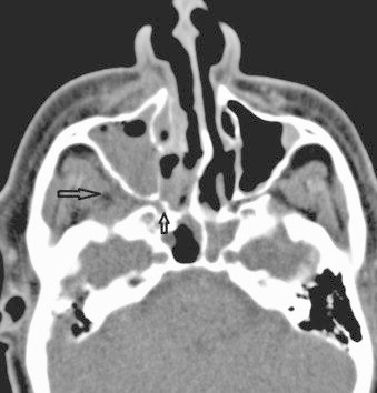
Pretreatment axial CT: note spread from the nasal cavity through the sphenopalatine foramen (small arrow) involving the right pterygopalatine fossa (big arrow). There is extension of infection into the right premaxillary soft tissues.
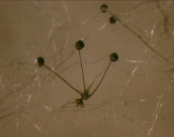
Rhizopus arrhizus.
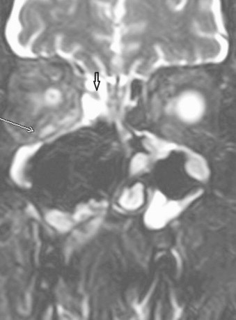
Coronal T2 FS MRI: black arrow shows a fungal concretion. White arrow reveals an enlargement and hyperintense signal of the right inferior rectus muscle and infiltration orbital fat.
References
-
- de Hoog GS, Guarro J, Gené J, Figueras MJ. Atlas of Clinical Fungo USB version. CBS-KNAW Fungal Biodiversity Centre and University Rovira i Virgili; 2014.
-
- Petrikkos G., Skiada A., Lortholary O., Roilides E., Walsh T.J., Kontoyiannis D.P. Epidemiology and clinical manifestations of mucormycosis. Clin Infect Dis. 2012;54(Suppl. 1):S23–S34. - PubMed
-
- Bonifaz A., Tirado-Sanchez A., Calderon L., Romero-Cabello R., Kassack J., Ponce R.M. Mucormycosis in children: a study of 22 cases in a Mexican hospital. Mycoses. 2014 - PubMed
-
- Kontoyiannis D.P., Lewis R.E. Invasive zygomycosis: update on pathogenesis, clinical manifestations, and management. Infect Dis Clin North Am. 2006;20(3):581–607. (vi) - PubMed
-
- Prasad K., Lalitha R.M., Reddy E.K., Ranganath K., Srinivas D.R., Singh J. Role of early diagnosis and multimodal treatment in rhinocerebral mucormycosis: experience of 4 cases. J Oral Maxillofac Surg. 2012;70(2):354–362. - PubMed
LinkOut - more resources
Full Text Sources
Other Literature Sources
