. 2015 Jan 22;517(7535):497-500.
doi: 10.1038/nature13896. Epub 2014 Nov 5.

mTORC1-mediated translational elongation limits intestinal tumour initiation and growth

Affiliations

mTORC1-mediated translational elongation limits intestinal tumour initiation and growth

William J Faller et al. Nature. .

Abstract

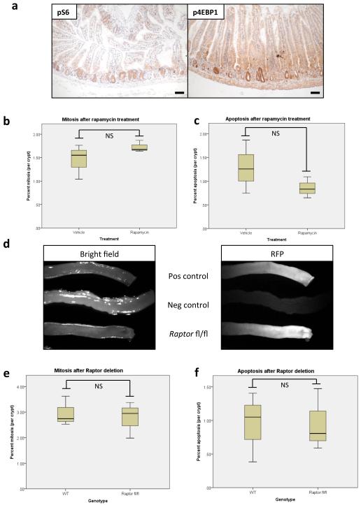
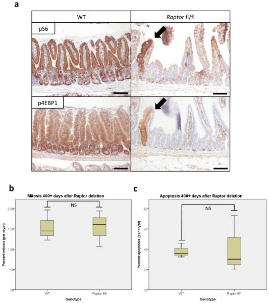
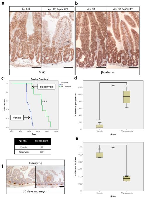
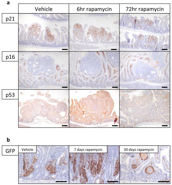
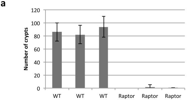
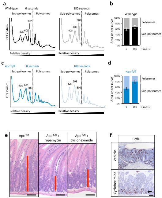
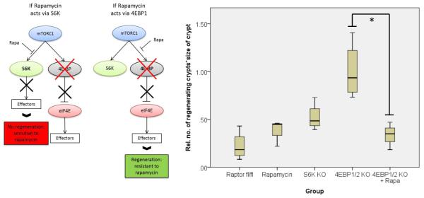
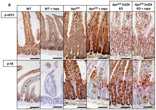
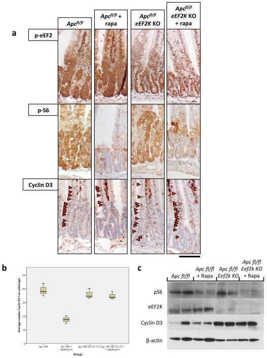
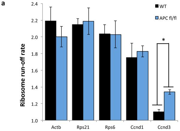
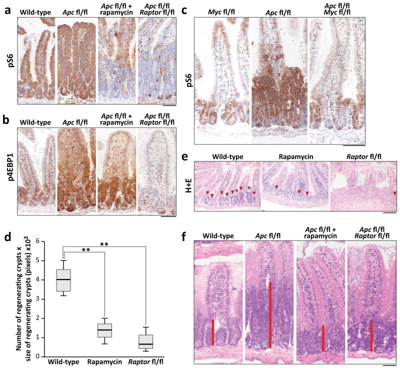
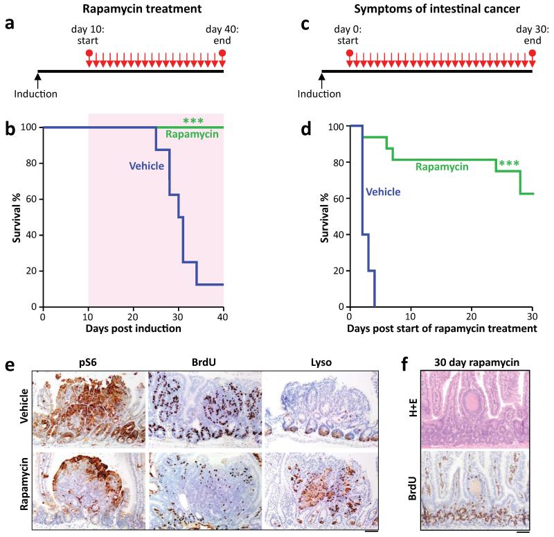
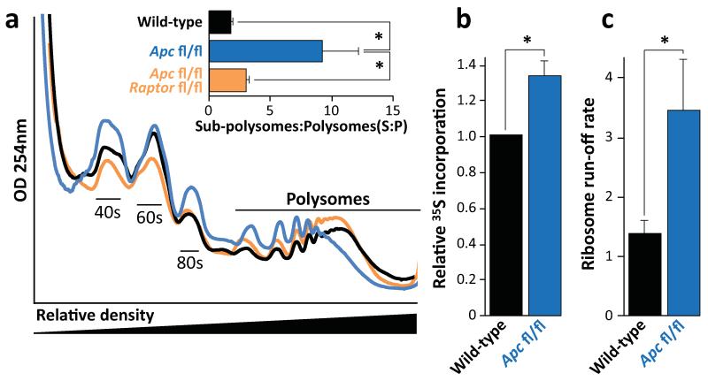
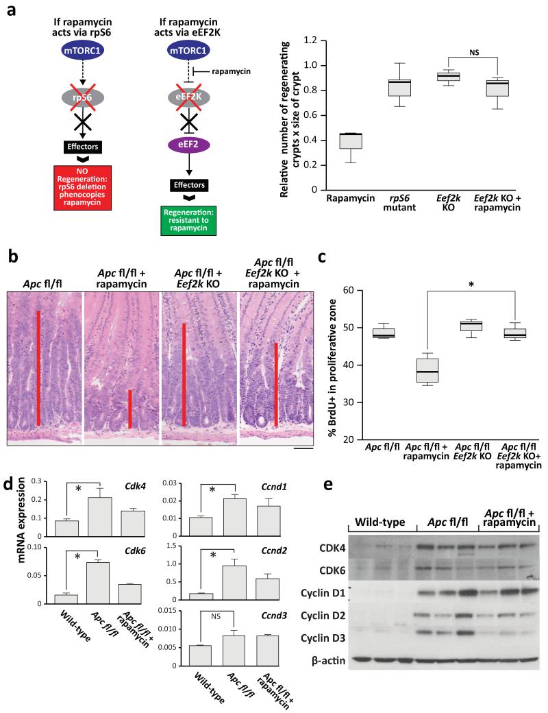
Inactivation of APC is a strongly predisposing event in the development of colorectal cancer, prompting the search for vulnerabilities specific to cells that have lost APC function. Signalling through the mTOR pathway is known to be required for epithelial cell proliferation and tumour growth, and the current paradigm suggests that a critical function of mTOR activity is to upregulate translational initiation through phosphorylation of 4EBP1 (refs 6, 7). This model predicts that the mTOR inhibitor rapamycin, which does not efficiently inhibit 4EBP1 (ref. 8), would be ineffective in limiting cancer progression in APC-deficient lesions. Here we show in mice that mTOR complex 1 (mTORC1) activity is absolutely required for the proliferation of Apc-deficient (but not wild-type) enterocytes, revealing an unexpected opportunity for therapeutic intervention. Although APC-deficient cells show the expected increases in protein synthesis, our study reveals that it is translation elongation, and not initiation, which is the rate-limiting component. Mechanistically, mTORC1-mediated inhibition of eEF2 kinase is required for the proliferation of APC-deficient cells. Importantly, treatment of established APC-deficient adenomas with rapamycin (which can target eEF2 through the mTORC1-S6K-eEF2K axis) causes tumour cells to undergo growth arrest and differentiation. Taken together, our data suggest that inhibition of translation elongation using existing, clinically approved drugs, such as the rapalogs, would provide clear therapeutic benefit for patients at high risk of developing colorectal cancer.


Figures

Extended Data Fig. 1
Extended Data Fig. 1. mTORC1 is activated following Wnt-signal and its inhibition does not affect homeostasis
a) Representative IHC of phospho-rpS6 and phospho-4EBP1 show mTORC1 activity during intestinal regeneration, 72hrs after 14Gy γ-irradiation (representative of 5 biological replicates); b+c) boxplots demonstrating that 72hrs of 10mg/kg rapamycin treatment does not alter mitosis or apoptosis in normal intestinal crypts. Whiskers are max and min, black line is median (n=4 per group; NS: not significant, Mann-Whitney U test); d) Intestines imaged on OV100 microscope, 96hrs post induction, for RFP. Tissue without the ROSA-tdRFP reporter (labelled Neg Control) show no RFP positivity, while the positive control and Raptor fl/fl intestines show high RFP positivity (representative of 3 biological replicates).; e+f) boxplot showing that Raptor deletion does not affect mitosis or apoptosis rates in intestinal crypts, 96hrs after induction. Whiskers are max and min, black line is median (n=4 per group; NS: not significant, Mann-Whitney U test). Scale bar = 100μm
Extended Data Fig. 2
Extended Data Fig. 2. Raptor deletion is maintained in the small intestine
a) Representative IHC of phospho-rpS6 and phospho-4EBP1 shows maintained loss of mTORC1 signalling 400+ days after Raptor deletion. Arrows indicate unrecombined escaper crypts that still show active mTORC1 signalling (Representative of 5 biological replicates).; b+c) boxplots showing that mitosis and apoptosis are unchanged 400+ days after Raptor deletion. Mitosis and apoptosis were counted on H+E sections and are quantified as percent mitosis or apoptosis per crypt. Whiskers are max and min, black line is median (n=5 per group; NS: not significant, Mann-Whitney U test). Scale bar = 100μm
Extended Data Fig. 3
Extended Data Fig. 3. Wnt-signalling is still active after Raptor deletion and Rapamycin treatment causes regression of established tumours
a+b) Representative IHC of MYC and β-catenin showing high MYC levels and nuclear localisation of β-catenin 96hrs after Apc and Apc/Raptor deletion, demonstrating active Wnt-signalling. Nuclear staining (as opposed to membranous staining) of β-catenin is indicative of active Wnt-signalling; Scale bar = 100μm (representative of 3 biological replicates). c) Kaplan-Meyer survival curve of ApcMin/+ mice treated with rapamycin when showing signs of intestinal neoplasia. 10mg/kg rapamycin treatment started when mice showed signs of intestinal disease, and was withdrawn after 30 days. Animals continued to be observed until signs of intestinal neoplasia. Death of animals in the rapamycin group almost always occurred following rapamycin withdrawal. (n=8 per group, ***p-value≤0.001, Log Rank test); d) boxplot showing that 72hr 10mg/kg rapamycin treatment causes an increase in Lysozyme positive cells in tumours. Percent Lysozyme positivity within tumours was calculated using Image J software (http://imagej.nih.gov/ij/). Whiskers are max and min, black line is median (10 tumours from each of 5 mice per group were measured, **p-value≤0.014, Mann-Whitney U test); e) boxplot showing that 72hrs 10mg/kg rapamycin treatment causes a decrease in BrdU positivity within tumours. Percent BrdU positivity within tumours was calculated using Image J software. Whiskers are max and min, black line is median (10 tumours from each of 5 mice per group were measured, **p-value≤0.021, Mann-Whitney U test); f) Representative IHC of Lysozyme, showing a lack of Lysozyme positive paneth cells in remaining cystic tumours after 30 days of 10mg/kg rapamycin treatment; Scale bar = 100μm (representative of 5 biological replicates).
Extended Data Fig. 4
Extended Data Fig. 4. IHC following rapamycin treatment
a) Representative IHC of p21, p16, and p53 after 6hrs and 72hrs of 10mg/kg rapamycin treatment. Staining shows no induction of these proteins in tumours following rapamycin treatment (representative of 5 biological replicates).; b) Representative IHC for LGR5 GFP showing high numbers of LGR5-positive cells after 7 and 30 days of 10mg/kg daily rapamycin treatment. (representative of 5 biological replicates). Scale bar = 100μm.
Extended Data Fig. 5
Extended Data Fig. 5. Raptor deletion in the intestinal crypt is lethal in vitro
a) Graph showing that Raptor deletion prevents intestinal crypts from growing ex vivo. Intestinal crypts were isolated and cultured as previously described, 96hrs after Cre induction. Number of viable organoids was counted by eye 72hrs after crypt isolation. Data are average ± s.d. (n=3 biological replicates per group)
Extended Data Fig 6
Extended Data Fig 6. Apc deletion increases translational elongation rates and Cycloheximide treatment phenocopies rapamycin treatment
a) Representative polysome profiles from wild-type ex vivo crypts incubated with harringtonine for 0 seconds (left) and 180 seconds (right) prior to harvest. (n=3 per time point) b) The areas under the sub-polysome (40S, 60S and 80S) and polysome sections as indicated by the dashed lines in a) were quantified and expressed as a percentage of their sum. Data in the bar graph is the average ± s.e.m (n=3 per time point). c) and d) show data for Apc deleted crypts as for wild-type in b) and c) (n=3 biological replicates). e) Representative H+E showing that 35mg/kg cycloheximide treatment phenocopies rapamycin treatment 96hrs after Apc deletion. Treatment began 24hrs after induction (n=3 biological replicates); f) Representative IHC for BrdU showing a loss of proliferation in tumours after 72hrs of 35mg/kg cycloheximide treatment. (n=3 biological replicates). Arrow highlights normal proliferating crypts. Scale bar = 100μm
Extended Data Fig. 7
Extended Data Fig. 7. S6k deletion decreases intestinal regeneration
Graphical representation of findings, and boxplot showing that murine intestinal regeneration following irradiation is dependent on S6K. Animals were exposed to 14Gy γ-irradiation, and intestinal regeneration was calculated 72hrs after exposure by counting the number of viable crypts and multiplying that by the average size of the regenerating crypts. Relative regeneration was calculated by comparing each group to WT regeneration. Rapamycin treatment arm is reproduced from Fig.4 for visual clarity. Whiskers are max and min, black line is median (n=6 per group, *p-value=0.034, Mann Whitney U test).
Extended Data Fig. 8
Extended Data Fig. 8. Eef2k deletion drives resistance to rapamycin
a) Representative IHC of phospho-eEF2 and phospho-S6 in WT, Apc-deficient and Apc- and Eef2k-deficient (with and without 72hrs 10mg/kg rapamycin treatment) shows that rapamycin is unable to induce eEF2 phosphorylation in the absence of eEF2K (n=6 biological replicates). Scale bar = 100μm
Extended Data Fig. 9
Extended Data Fig. 9. Cyclin D3 is regulated at the level of elongation
a) Representative IHC of Apcfl/fl intestines with and without Eef2k deletion. Antibodies to eEF2K, phospho-S6, and Cyclin D3 are shown (representative of 3 biological replicates). Following Eef2k KO, Cyclin D3 levels are no longer decreased upon 10mg/kg rapamycin treatment; b) boxplot showing the number of Cyclin D3 positive cells per crypt, 96hrs after Apc deletion, with and without 10mg/kg rapamycin treatment. Graph shows that in Eef2k KO animals, rapamycin no longer reduced Cyclin D3 levels (n=3 biological replicates per group; *p-value≤0.05, Mann Whitney U test); c) western blot analysis of intestinal epithelial cells from Apcfl/fl and Apcfl/fl Eef2k KO, with and without 10mg/kg rapamycin. Antibodies to eEF2K, phospho-S6, Cyclin D3 and β-actin are shown. Each well represents a different mouse from the relevant group. Cyclin D3 levels are no longer reduced following Eef2k deletion. Scale bar = 100μm.
Extended Data Fig. 10
Extended Data Fig. 10. Ribosomes elongate faster on Ccnd3 following Apc deletion
The ribosome run-off rate of various messages was measured as in Figure 3. Elongation of Ccnd3 was significantly increased, while Actb, Rps21, Rps6 and Ccnd1 remain unchanged. Data are average ± s.e.m. (n=3 biological replicates per group; *p-value≤0.05, Mann Whitney U test).
Figure 1
Figure 1. mTORC1 is essential for Wnt-driven proliferation in a MYC-dependent manner
a+b) Representative IHC of phospho-rpS6 and phospho-4EBP1 showing increased staining 96hrs following Apc deletion. Raptor deletion caused a loss of positivity in both, whereas 10mg/kg rapamycin treatment (beginning at 24hrs) specifically disrupts rpS6 phosphorylation (representative of 6 biological replicates); c) Representative IHC of phospho-rpS6 96hrs after Cre induction showing that Wnt-driven rpS6 phosphorylation is MYC dependent (representative of 3 biological replicates); d) Animals were exposed to 14Gy γ-irradiation and intestinal regeneration was measured 72hrs later, by counting the number of viable crypts and multiplying that by the average size of the regenerating crypts. Boxplot shows that 10mg/kg rapamycin treatment and Raptor deletion significantly decrease intestinal regeneration. Whiskers are max and min, black line is median (n=5 biological replicates per group; **p-value<0.02, Mann-Whitney U test); e) Representative H+E staining of regenerating intestines 72hrs after exposure to 14Gy γ-irradiation. Arrowheads indicate regenerating crypt; (representative of 5 biological replicates).; f) Representative H+E staining 96hrs after Apc loss, showing that 10mg/kg rapamycin treatment or Raptor deletion prevent Wnt-driven proliferation (representative of 6 biological replicates). Treatment began 24hrs after Apc deletion. Red bar is graphical representation of crypt size. Scale bar = 100μm.
Figure 2
Figure 2. Apc-driven tumourigenesis requires mTORC1 activation
a+b) Graphical representation of prophylactic rapamycin treatment strategy and Kaplan-Meyer survival curve showing that prophylactic rapamycin treatment prevents tumourigenesis. 10mg/kg rapamycin treatment began at Day 10 post-Apc deletion, and lasted 30 days, after which mice were sampled. Area highlighted by red indicates duration of rapamycin treatment (n=8, vehicle; n=13 rapamycin; ***p-value≤0.001, Log Rank test); c+d) Graphical representation of chemotherapeutic rapamycin treatment strategy and Kaplan-Meyer survival curve showing that rapamycin treatment can regress established intestinal tumours. 10mg/kg rapamycin treatment started when mice showed signs of intestinal disease, and lasted 30 days, after which mice were sampled. Graph represents survival while on rapamycin treatment (n=5, vehicle; n= 16, rapamycin; ***p-value≤0.001, Log Rank test); e) Representative IHC of phospho-rpS6, BrdU and Lysozyme, showing that 72hrs of 10mg/kg rapamycin treatment causes a loss in rpS6 phosphorylation and BrdU positivity, and an increase in lysozyme staining in intestinal tumours (representative of 5 biological replicates); f) Representative H+E and IHC for BrdU showing that small, non-proliferative lesions remain after 30 days of 10mg/kg rapamycin treatment (representative of 5 biological replicates). Scale bar = 100μm
Figure 3
Figure 3. mTORC1 drives increased translational elongation
a) Representative polysome profiles of intestinal epithelial cells showing altered RNA distribution 96hrs after Apc deletion. Bar graph represents the ratio of sub-polysomes compared to polysomes (S:P). Data are average ± s.e.m. (n=3 per group; *p-value≤0.05, Mann-Whitney U test); b) Intestinal crypt culture was pulsed for 30 min with 35S-labelled methionine/cysteine. Incorporation of 35S into protein was quantified by scintillation counting and normalized to total protein. Apc deletion increases 35S incorporation. Data are average ± s.e.m. (n=3 biological replicates per group; *p-value≤0.05, Mann-Whitney U test); c) The ribosome run-off rate was measured by addition of the initiation inhibitor harringtonine to ex vivo crypts from wild-type and Apc deleted mice. Harringtonine was added for 0 or 180 seconds and the increase in sub-polysomes (S) relative to polysome (P) calculated. This run-off rate represents the shift in S:P between the two time points, which is proportional to elongation speed. Data are average ± s.e.m. (n=3 biological replicates per group; *p-value≤0.05, Mann Whitney U test). Also see Supplemental Figure 7.
Figure 4
Figure 4. mTORC1 signalling via eEF2K controls intestinal proliferation following Wnt-signalling
a) Graphical representation of findings and boxplots showing that murine intestinal regeneration following irradiation implicates the mTORC1-S6K-eEF2K axis in Wnt-driven proliferation. Animals were exposed to 14Gy γ-irradiation, and intestinal regeneration was calculated 72hrs after exposure, by examining the number and size of regenerating crypts, relative to WT regenerating intestines. Whiskers are max and min, black line is median (n=6 per group; *p-value<0.05, NS: not significant, Mann-Whitney U test); b+c) Representative H+E and boxplot showing Eef2k deletion confers resistance to 10mg/kg rapamycin treatment, 96hrs after Apc deletion. Treatment began 24hrs after induction. Red bar is graphical representation of crypt size. Whiskers are max and min, black line is median (n=3 biological replicates per group; *p-value<0.05, Mann-Whitney U test); d) qRT-PCR of intestinal epithelial cells using primers for Cdk4, Cdk6, Ccnd1, Ccnd2 and Ccnd3. Ccnd3 is not regulated at the transcriptional level. Data were normalised to Gapdh. Data are average ± s.e.m. (n=3 biological replicates per group, *p-value≤0.05, NS: not significant, Mann Whitney U test); e) Western blot analysis of intestinal epithelial cells from each group. Antibodies to CDK4, CDK6, Cyclin D1, Cyclin D2, Cyclin D3 and β-actin are shown. Each well represents a different mouse from the relevant group, and are the same samples used for the qRT-PCR. Scale bar = 100μm.

Comment in

References

    1. Kinzler KW, Vogelstein B. Lessons from hereditary colorectal cancer. Cell. 1996;87:159–170. - PubMed
    1. Korinek V, et al. Constitutive transcriptional activation by a beta-catenin-Tcf complex in APC−/− colon carcinoma. Science. 1997;275:1784–1787. - PubMed
    1. Ashton GH, et al. Focal adhesion kinase is required for intestinal regeneration and tumorigenesis downstream of Wnt/c-Myc signaling. Developmental cell. 2010;19:259–269. doi:10.1016/j.devcel.2010.07.015. - PMC - PubMed
    1. Fujishita T, Aoki K, Lane HA, Aoki M, Taketo MM. Inhibition of the mTORC1 pathway suppresses intestinal polyp formation and reduces mortality in ApcDelta716 mice. Proceedings of the National Academy of Sciences of the United States of America. 2008;105:13544–13549. doi:10.1073/pnas.0800041105. - PMC - PubMed
    1. Gulhati P, et al. Targeted inhibition of mammalian target of rapamycin signaling inhibits tumorigenesis of colorectal cancer. Clin Cancer Res. 2009;15:7207–7216. doi:10.1158/1078-0432.CCR-09-1249. - PMC - PubMed

Methods Reference

    1. El Marjou F, et al. Tissue-specific and inducible Cre-mediated recombination in the gut epithelium. Genesis. 2004;39:186–193. doi:10.1002/gene.20042. - PubMed
    1. Barker N, et al. Identification of stem cells in small intestine and colon by marker gene Lgr5. Nature. 2007;449:1003–1007. doi:nature06196 [pii] 10.1038/nature06196. - PubMed
    1. Shibata H, et al. Rapid colorectal adenoma formation initiated by conditional targeting of the Apc gene. Science. 1997;278:120–123. - PubMed
    1. Moser AR, Pitot HC, Dove WF. A dominant mutation that predisposes to multiple intestinal neoplasia in the mouse. Science. 1990;247:322–324. - PubMed
    1. de Alboran IM, et al. Analysis of C-MYC function in normal cells via conditional gene-targeted mutation. Immunity. 2001;14:45–55. - PubMed

Publication types

MeSH terms

Substances