

Reference values for healthy human myocardium using a T1 mapping methodology: results from the International T1 Multicenter cardiovascular magnetic resonance study
- PMID: 25384607
- PMCID: PMC4203908
- DOI: 10.1186/s12968-014-0069-x
Reference values for healthy human myocardium using a T1 mapping methodology: results from the International T1 Multicenter cardiovascular magnetic resonance study
Abstract
Background: T1 mapping is a robust and highly reproducible application to quantify myocardial relaxation of longitudinal magnetisation. Available T1 mapping methods are presently site and vendor specific, with variable accuracy and precision of T1 values between the systems and sequences. We assessed the transferability of a T1 mapping method and determined the reference values of healthy human myocardium in a multicenter setting.
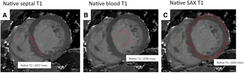
Methods: Healthy subjects (n=102; mean age 41 years (range 17-83), male, n=53 (52%)), with no previous medical history, and normotensive low risk subjects (n=113) referred for clinical cardiovascular magnetic resonance (CMR) were examined. Further inclusion criteria for all were absence of regular medication and subsequently normal findings of routine CMR. All subjects underwent T1 mapping using a uniform imaging set-up (modified Look- Locker inversion recovery, MOLLI, using scheme 3(3)3(3)5)) on 1.5 Tesla (T) and 3 T Philips scanners. Native T1-maps were acquired in a single midventricular short axis slice and repeated 20 minutes following gadobutrol. Reference values were obtained for native T1 and gadolinium-based partition coefficients, λ and extracellular volume fraction (ECV) in a core lab using standardized postprocessing.
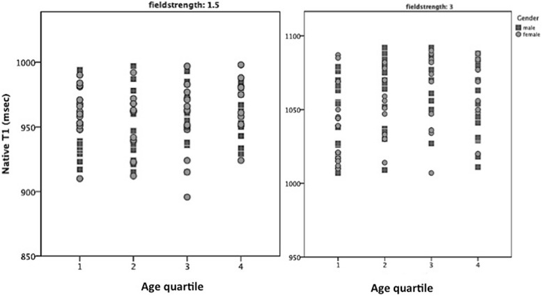
Results: In healthy controls, mean native T1 values were 950±21 msec at 1.5 T and 1052±23 at 3 T. λ and ECV values were 0.44±0.06 and 0.25±0.04 at 1.5 T, and 0.44±0.07 and 0.26±0.04 at 3 T, respectively. There were no significant differences between healthy controls and low risk subjects in routine CMR parameters and T1 values. The entire cohort showed no correlation between age, gender and native T1. Cross-center comparisons of mean values showed no significant difference for any of the T1 indices at any field strength. There were considerable regional differences in segmental T1 values. λ and ECV were found to be dose dependent. There was excellent inter- and intraobserver reproducibility for measurement of native septal T1.
Conclusion: We show transferability for a unifying T1 mapping methodology in a multicenter setting. We provide reference ranges for T1 values in healthy human myocardium, which can be applied across participating sites.
Figures


References
-
- Kwon DH, Halley CM, Carrigan TP, Zysek V, Popovic ZB, Setser R, Schoenhagen P, Starling RC, Flamm SD, Desai MY. Extent of left ventricular scar predicts outcomes in ischemic cardiomyopathy patients with significantly reduced systolic function: a delayed hyperenhancement cardiac magnetic resonance study. JACC Cardiovasc Imaging. 2009;2(1):34–44. doi: 10.1016/j.jcmg.2008.09.010. - DOI - PubMed
-
- Schelbert EB, Cao JJ, Sigurdsson S, Aspelund T, Kellman P, Aletras AH, Dyke CK, Thorgeirsson G, Eiriksdottir G, Launer LJ, Gudnason V, Harris TB, Arai AE. Prevalence and prognosis of unrecognized myocardial infarction determined by cardiac magnetic resonance in older adults. JAMA. 2012;308(9):890–896. doi: 10.1001/2012.jama.11089. - DOI - PMC - PubMed
-
- Neilan TG, Coelho-Filho OR, Danik SB, Shah RV, Dodson JA, Verdini DJ, Tokuda M, Daly CA, Tedrow UB, Stevenson WG, Jerosch-Herold M, Ghoshhajra BB, Kwong RY. CMR quantification of myocardial scar provides additive prognostic information in nonischemic cardiomyopathy. JACC Cardiovasc Imaging. 2013;6(9):944–954. doi: 10.1016/j.jcmg.2013.05.013. - DOI - PMC - PubMed
-
- Gulati A, Jabbour A, Ismail TF, Guha K, Khwaja J, Raza S, Morarji K, Brown TD, Ismail NA, Dweck MR, Di Pietro E, Roughton M, Wage R, Daryani Y, O’Hanlon R, Sheppard MN, Alpendurada F, Lyon AR, Cook SA, Cowie MR, Assomull RG, Pennell DJ, Prasad SK. Association of fibrosis with mortality and sudden cardiac death in patients with nonischemic dilated cardiomyopathy. JAMA. 2013;309(9):896–908. doi: 10.1001/jama.2013.1363. - DOI - PubMed
-
- Iles L, Pfluger H, Lefkovits L, Butler MJ, Kistler PM, Kaye DM, Taylor AJ. Myocardial fibrosis predicts appropriate device therapy in patients with implantable cardioverter-defibrillators for primary prevention of sudden cardiac death. J Am Coll Cardiol. 2011;15(57(7)):821–828. doi: 10.1016/j.jacc.2010.06.062. - DOI - PubMed
Publication types
MeSH terms
Substances
Grants and funding
LinkOut - more resources
Full Text Sources
Other Literature Sources
Medical
Miscellaneous
