. 2014 Dec 15;592(24):5493-505.
doi: 10.1113/jphysiol.2014.281253. Epub 2014 Nov 10.

The effects of dexamethasone on post-asphyxial cerebral oxygenation in the preterm fetal sheep

Affiliations

The effects of dexamethasone on post-asphyxial cerebral oxygenation in the preterm fetal sheep

Christopher A Lear et al. J Physiol. .

Abstract

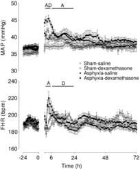
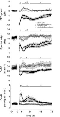
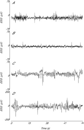
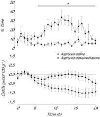
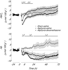
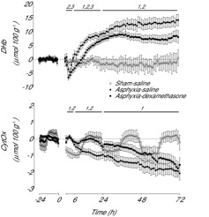
Exposure to clinical doses of the glucocorticoid dexamethasone increases brain activity and causes seizures in normoxic preterm fetal sheep without causing brain injury. In contrast, the same treatment after asphyxia increased brain injury. We hypothesised that increased injury was in part mediated by a mismatch between oxygen demand and oxygen supply. In preterm fetal sheep at 0.7 gestation we measured cerebral oxygenation using near-infrared spectroscopy, electroencephalographic (EEG) activity, and carotid blood flow (CaBF) from 24 h before until 72 h after asphyxia induced by 25 min of umbilical cord occlusion. Ewes received dexamethasone intramuscularly (12 mg 3 ml(-1)) or saline 15 min after the end of asphyxia. Fetuses were studied for 3 days after occlusion. During the first 6 h of recovery after asphyxia, dexamethasone treatment was associated with a significantly greater fall in CaBF (P < 0.05), increased carotid vascular resistance (P < 0.001) and a greater fall in cerebral oxygenation as measured by the difference between oxygenated and deoxygenated haemoglobin (delta haemoglobin; P < 0.05). EEG activity was similarly suppressed in both groups. From 6 to 10 h onward, dexamethasone treatment was associated with a return of CaBF to saline control levels, increased EEG power (P < 0.005), greater epileptiform transient activity (P < 0.001), increased oxidised cytochrome oxidase (P < 0.05) and an attenuated increase in [delta haemoglobin] (P < 0.05). In conclusion, dexamethasone treatment after asphyxia is associated with greater hypoperfusion in the critical latent phase, leading to impaired intracerebral oxygenation that may exacerbate neural injury after asphyxia.


Figures

Figure 1
Figure 1. Time sequence of changes in MAP (mmHg) and FHR (bpm) from 24 h before until 72 h after umbilical cord occlusion in sham–saline (n = 13), sham–dexamethasone (n = 12), asphyxia–saline (n = 18) and asphyxia–dexamethasone (n = 15) groups
Data are 1 h means ± SEM. A, effect of asphyxia, P < 0.05; D, effect of dexamethasone treatment, P < 0.05; FHR, fetal heart rate; MAP, mean arterial pressure.
Figure 2
Figure 2. Time sequence of changes in EEG power (dB, change from baseline), spectral edge frequency (Hz), CaBF (ml min−1) and CaVR (mmHg ml−1min−1) from 24 h before until 72 h after umbilical cord occlusion in sham–saline (n = 13), sham–dexamethasone (n = 12), asphyxia–saline (n = 18) and asphyxia–dexamethasone (n = 15) groups
Data are 1 h means ± SEM. A, effect of asphyxia, P < 0.05; CaBF, carotid blood flow; CaVR, carotid vascular resistance; D, effect of dexamethasone treatment, P < 0.05.
Figure 3
Figure 3. Examples of raw EEG activity from sham–saline controls and after asphyxia and dexamethasone treatment
A, example of the normal discontinuous, mixed amplitude and frequency EEG activity from the sham–saline group. B, example of post-asphyxial EEG activity 4 h after asphyxia in the asphyxia–saline group, showing suppressed background activity interspersed with the presence of sharp and fast wave transients. C, effect of dexamethasone on post-asphyxial EEG activity 4 h after asphyxia in the asphyxia–dexamethasone group, showing markedly higher background amplitude and the presence of prolonged abnormal slow wave rolling activity. D, effect of dexamethasone on normal EEG activity in the sham–dexamethasone group, showing the presence of similar low frequency, high amplitude activity after combined asphyxia and dexamethasone treatment. Note the different scale used in (D).
Figure 4
Figure 4. Time sequence of changes in percentage time per hour of prolonged slow wave interictal activity and levels of oxidised CytOx (μmol 100 g−1) for the first 24 h after umbilical cord occlusion in asphyxia–saline and asphyxia–dexamethasone groups
Data are 1 h means ± SEM. *Asphyxia–saline vs. asphyxia–dexamethasone, P < 0.05. CytOx, cytochrome oxidase.
Figure 5
Figure 5. Time sequence of changes in Hb (μmol 100 g−1) and HbO2 (μmol 100 g−1) from 24 h before until 72 h after umbilical cord occlusion in sham–saline (n = 5), asphyxia–saline (n = 9) and asphyxia–dexamethasone (n = 7) groups
Data are 1 h means ± SEM. 1, asphyxia–saline vs. sham–saline, P < 0.05; 2, asphyxia–dexamethasone vs. sham–saline, P < 0.05; 3, asphyxia–saline vs. asphyxia–dexamethasone, P < 0.05; Hb, deoxyhaemoglobin; HbO2, oxyhaemoglobin.
Figure 6
Figure 6. Time sequence of changes in DHb (μmol 100 g−1) and levels of oxidised CytOx (μmol 100 g−1) from 24 h before until 72 h after umbilical cord occlusion in sham–saline (n = 5), asphyxia–saline (n = 9) and asphyxia–dexamethasone (n = 7) groups
Data are 1 h means ± SEM. 1, Asphyxia–saline vs. sham–saline, P < 0.05; 2, asphyxia–dexamethasone vs. sham–saline, P < 0.05; 3, asphyxia–saline vs. asphyxia–dexamethasone, P < 0.05; CytOx, cytochrome oxidase; DHb, delta haemoglobin.

References

    1. Abraham I, Juhasz G, Kekesi KA, Kovacs KJ. Effect of intrahippocampal dexamethasone on the levels of amino acid transmitters and neuronal excitability. Brain Res. 1996;733:56–63. - PubMed
    1. Bennet L, Booth LC, Drury PP, Quaedackers JS, Gunn AJ. Preterm neonatal cardiovascular instability: does understanding the fetus help evaluate the newborn. Clin Exp Pharmacol Physiol. 2012a;39:965–972. - PubMed
    1. Bennet L, Booth L, Gunn AJ. Potential biomarkers for hypoxic-ischemic encephalopathy. Semin Fetal Neonatal Med. 2010;15:253–260. - PMC - PubMed
    1. Bennet L, Davidson JO, Koome M, Gunn AJ. Glucocorticoids and preterm hypoxic-ischemic brain injury: the good and the bad. J Pregnancy. 2012b;2012:751694. - PMC - PubMed
    1. Bennet L, Kozuma S, McGarrigle HHG, Hanson MA. Temporal changes in fetal cardiovascular, behavioural, metabolic and endocrine responses to maternally administered dexamethasone in the late gestation fetal sheep. Br J Obstet Gynaecol. 1999a;106:331–339. - PubMed

Publication types

MeSH terms

LinkOut - more resources