Comparative Study
. 2014;36(6):9728.
doi: 10.1007/s11357-014-9728-y. Epub 2014 Nov 11.

A comprehensive study of myocardial redox homeostasis in naturally and mimetically aged rats

Affiliations
Comparative Study

A comprehensive study of myocardial redox homeostasis in naturally and mimetically aged rats

Tamer Cebe et al. Age (Dordr). 2014.

Abstract

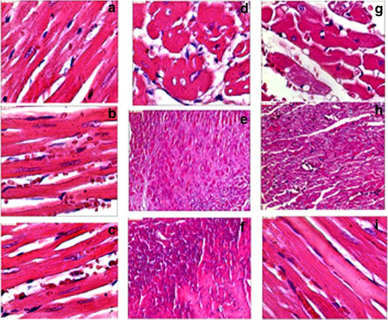
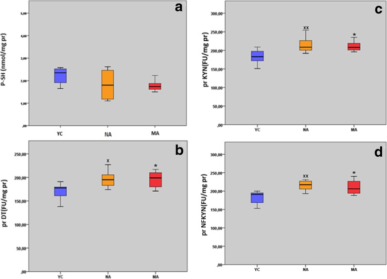
Age-related myocardial dysfunction has important implications with impaired redox homeostasis. Current study focused on investigation of redox homeostasis and histopathological changes in the myocardium of mimetically (MA), naturally aged (NA), and young control (YC) rats. Chronic D-galactose administration to young male Wistar rats (5 months old) was used to set up experimental aging models. We investigated 16 different oxidative damage biomarkers which have evaluated redox homeostasis of cellular macromolecules such as protein, lipid, and DNA. As a protein oxidation biomarker, advanced oxidation end products, protein carbonyl groups, protein-bound advanced glycation end products, dityrosine, kynurenine, and N-formylkynurenine concentrations in MA and NA rats were found to be significantly higher compared to those in YC rats. On the other hand, the levels of protein thiol groups were not significantly different between groups, whereas lipid peroxidation biomarkers such as conjugated diens, lipid hydroperoxides, and malondialdehyde in MA and NA rats were found to be significantly higher in comparison to those in YCs. For the assessment of oxidative DNA damage, we analyzed eight hydroxy-5'-deoxyguanosine concentrations of MA and NA groups which were higher than YCs. As an antioxidant status in the MA and NA groups, Cu-Zn superoxide dismutase, ferric reducing antioxidant power, and total thiol levels were lower than those in the YCs. Only nonprotein thiol levels were not significantly different. We also observed similar histopathological changes in MA and NA rats. We concluded that the mimetic aging model could be considered as a reliable experimental model for myocardial senescence.


Figures

Fig. 1
Fig. 1
Histological evaluation of the myocardial tissue of chronologically and mimetically aged rats and their young controls. Histological evaluation of the myocardial tissue of young control rats. ac Branching muscle fibers can be observed. Centrally localized nuclei and cross striation can also be seen very clearly (H&E ×40). Connective tissue rich in capillaries can be seen between fibers (H&E ×40). Histological evaluation of the myocardial tissue of chronologically aged rats. d Cell cytoplasms have lots of vacuoles in cross section of the cardiac myocytes. Prominent intercellular edema is present, as well (H&E ×40). e Muscle fibers are replaced by spreading connective tissue (H&E ×40). f Degenerated muscle fibers with no visible myofilaments cover a wide area (HE ×40). Histological evaluation of the myocardial tissue of mimetically aged rats. g Cell cytoplasms have lots of vacuoles in cross section of the cardiac myocytes. Intercellular edema is attracting attention, as well. Cytoplasmic loss of myofibers is noticed (HE ×40). h Randomly and sparsely distributed myofibers are giving their places into connective tissue (HE ×10). i Degenerated, pale myofibers accompanied with disappearance of myofilaments are seen (H&E ×40)
Fig. 2
Fig. 2
General protein oxidation biomarkers in myocardial tissue of naturally aged rats (NA; n = 8), mimetically aged rats (MA; n = 8), and their corresponding young controls (YC; n = 8). Results were expressed as mean ± SEM. Data were statistically different between groups (*p < 0.05; **p < 0.01; YC vs MA) (x p < 0.05; xx p < 0.01; YC vs NA). PCO protein carbonyl groups, AOPP advanced oxidation protein end products, prAGEs protein-bound advanced glycation end products
Fig. 3
Fig. 3
Specific protein oxidation biomarkers in the myocardial tissue of naturally aged rats (NA; n = 8), mimetically aged rats (MA; n = 8), and their corresponding young controls (YC; n = 8). Results were expressed as mean ± SEM. Data were statistically different between groups (*p < 0.05; YC vs MA) (x p < 0.05; xx p < 0.01; YC vs NA). P-SH protein thiol, prDT protein-bound dityrosine, prKYN protein-bound kynurenine, prN-FKYN protein-bound N′-formylkynurenine
Fig. 4
Fig. 4
Lipid peroxidation biomarkers in myocardial tissue of naturally aged rats (NA; n = 8), mimetically aged rats (MA; n = 8), and their corresponding young controls (YC; n = 8). Results were expressed as mean ± SEM. Data were statistically different between groups (*p < 0.05; ***p < 0.001; YC vs MA) (xx p < 0.01; xxx p < 0.001; YC vs NA). MDA malondialdehyde, L-OOHs lipid hydroperoxides, ttCDs trans-trans conjugated diens, ctCDs cis–trans-conjugated diens
Fig. 5
Fig. 5
8-Hydroxy-2′-deoxyguanosine concentrations of the myocardial tissue of naturally aged rats (NA; n = 8), mimetically aged rats (MA; n = 8), and their corresponding young controls (YC; n = 8). Results were expressed as mean ± SEM. Data were statistically different between groups (***p < 0.001; YC vs MA) (xxx p < 0.001; YC vs NA). 8-OHdG 8-hydroxy-2′-deoxyguanosine
Fig. 6
Fig. 6
Antioxidant capacity biomarkers in myocardial tissue of naturally aged rats (NA; n = 8), mimetically aged rats (MA; n = 8), and their corresponding young controls (YC; n = 8). Results are expressed as mean ± SEM. Data were statistically different between groups (**p < 0.01; ***p < 0.001; YC vs MA) (x p < 0.05; xx p < 0.01; xxx p < 0.001; YC vs NA). Cu–Zn SOD Cu–Zn superoxide dismutase, FRAP ferric reducing antioxidant power, T-SH total thiol groups, NP-SH nonprotein thiol groups

References

    1. Aydın S, Yanar K, Atukeren P, Dalo E, Sitar ME, Uslu E, Caf N, Çakatay U. Comparison of oxidative stress biomarkers in renal tissues of D-galactose induced, naturally aged and young rats. Biogerontology. 2012;13:251–260. doi: 10.1007/s10522-011-9370-3. - DOI - PubMed
    1. Babusıkova E, Kaplan P, Lehotsky J, Jesenak M, Dobrota D. Oxidative modification of rat cardiac mitochondrial membranes and myofibrils by hydroxyl radicals. Gen Physiol Biophys. 2004;23:327–335. - PubMed
    1. Babusıkova E, Jesenak M, Dobrota D, Tribulova N, Kaplan P. Age-dependent effect of oxidative stress on cardiac sarcoplasmic reticulum vesicles. Physiol Res. 2008;57:S49–S54. - PubMed
    1. Baraibar MA, Liu L, Ahmed EK, Friguet B. Protein oxidative damage at the crossroads of cellular senescence, aging, and age-related diseases. Oxidative Med Cell Longev. 2012;2012:919832. doi: 10.1155/2012/919832. - DOI - PMC - PubMed
    1. Benzi IF, Strain JJ. Ferric reducing/antioxidant power assay: direct measure of total antioxidant activity of biological fluids and modified version for simultaneous measurement of total antioxidant power and ascorbic acid concentration. Methods Enzymol. 1999;299:15–27. doi: 10.1016/S0076-6879(99)99005-5. - DOI - PubMed

Publication types