. 2015 Jan 2;290(1):25-34.
doi: 10.1074/jbc.M114.609156. Epub 2014 Nov 10.

Human CHD2 is a chromatin assembly ATPase regulated by its chromo- and DNA-binding domains

Affiliations

Human CHD2 is a chromatin assembly ATPase regulated by its chromo- and DNA-binding domains

Jessica C Liu et al. J Biol Chem. .

Abstract

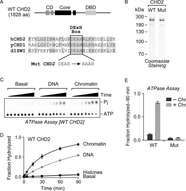
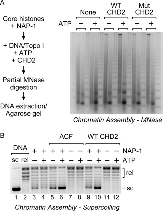
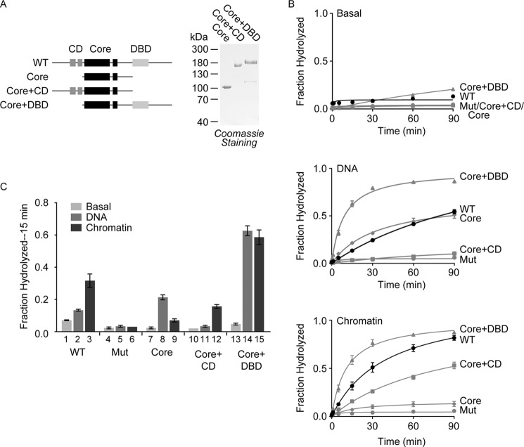
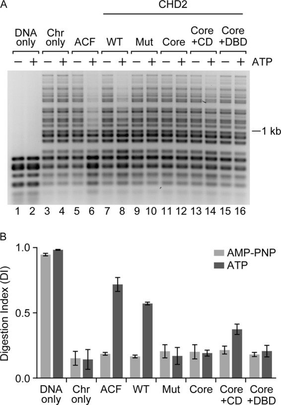
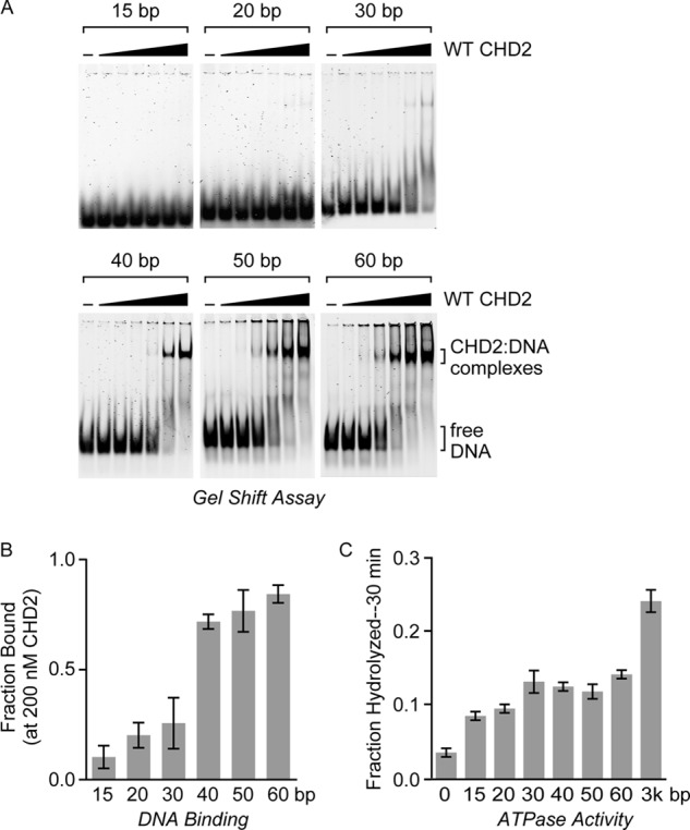
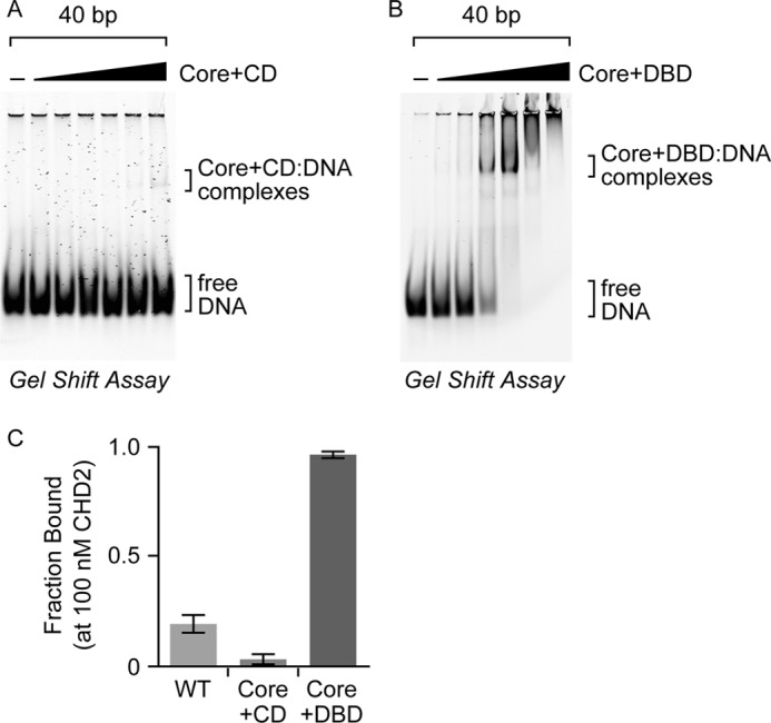
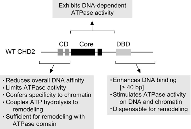
Chromodomain helicase DNA-binding protein 2 (CHD2) is an ATPase and a member of the SNF2-like family of helicase-related enzymes. Although deletions of CHD2 have been linked to developmental defects in mice and epileptic disorders in humans, little is known about its biochemical and cellular activities. In this study, we investigate the ATP-dependent activity of CHD2 and show that CHD2 catalyzes the assembly of chromatin into periodic arrays. We also show that the N-terminal region of CHD2, which contains tandem chromodomains, serves an auto-inhibitory role in both the DNA-binding and ATPase activities of CHD2. While loss of the N-terminal region leads to enhanced chromatin-stimulated ATPase activity, the N-terminal region is required for ATP-dependent chromatin remodeling by CHD2. In contrast, the C-terminal region, which contains a putative DNA-binding domain, selectively senses double-stranded DNA of at least 40 base pairs in length and enhances the ATPase and chromatin remodeling activities of CHD2. Our study shows that the accessory domains of CHD2 play central roles in both regulating the ATPase domain and conferring selectivity to chromatin substrates.

Keywords: ATPase; CHD2; Chromatin Remodeling; Chromodomains; DNA-binding Protein; Epilepsy; Nucleosome.


Figures

FIGURE 1.
FIGURE 1.
WT CHD2 is a chromatin-stimulated ATPase. A, top, the WT human CHD2 protein contains a central SNF2-like ATPase domain (Core) that is flanked by tandem CDs and a putative DBD. Bottom, a partial alignment of human CHD2 (hCHD2), yeast CHD1 (yCHD1), and Drosophila ISWI (dISWI) highlights the conserved DEXH sequence of the Walker B box. We used site-directed mutagenesis to clone a mutant version of CHD2 (Mut) that contains a two-amino acid alanine substitution of the Asp-617 and Glu-618 residues. B, WT and Mut CHD2 were purified from baculovirus-infected cells and analyzed by SDS-PAGE and Coomassie staining. C, a representative radiometric ATPase assay used to measure the ability of CHD2 to hydrolyze ATP over time. ATPase reactions with WT CHD2 alone (Basal) or containing DNA or chromatin were incubated for 0, 0.5, 1, 5, 15, 30, 60, or 90 min, stopped by the addition of EDTA, and resolved by TLC on PEI-cellulose plates. The positions of the ATP and released phosphate on the TLC plate are indicated. D, quantification of the ATPase assays with WT CHD2 stimulated by chromatin, DNA, or core histones. E, quantification of the radiometric ATPase assays using purified WT or Mut CHD2 protein in the presence or absence of chromatin. The fraction of ATP hydrolyzed was calculated and the values shown are mean and S.D.; n = 3.
FIGURE 2.
FIGURE 2.
CHD2 catalyzes the assembly of periodic nucleosome arrays. A, left, nucleosome assembly reactions were performed to determine whether CHD2 assembles periodic nucleosome arrays. Core histones (1.4 μg) were pre-incubated with the histone chaperone NAP-1 (13 μg). Naked plasmid DNA (1.4 μg), Topo I, and WT or Mut CHD2 (100 nm) was then added in the presence or absence of ATP. The reactions were then partially digested with a low (left-hand lane) or a high (right-hand lane) concentration of MNase. The digested DNA was precipitated and analyzed by agarose gel electrophoresis and EtBr-staining. A 123-bp repeat marker was run between each pair of samples. Right, the assembly of periodic nucleosome arrays was analyzed by partial MNase digestion. A representative agarose gel showing the presence of a DNA ladder formed upon assembly of nucleosomes by CHD2 and ATP. B, supercoiling analysis of chromatin assembly reactions. Core histones (0.35 μg) were pre-incubated with the histone chaperone NAP-1 (2 μg). Naked plasmid DNA (0.35 μg), Topo I, and ACF (20 nm) or WT CHD2 (20 nm) was then added in the presence or absence of ATP (AMP-PNP used as negative ATP control). The plasmid DNA was then precipitated and analyzed by agarose gel electrophoresis and EtBr-staining. sc, supercoiled; rel, relaxed.
FIGURE 3.
FIGURE 3.
The accessory domains of CHD2 regulate the core ATPase domain. A, left, a series of CHD2 deletion proteins was generated that include removal of the C-terminal region containing the putative DNA-binding domain (Core+CD), removal of the N-terminal region containing the tandem chromodomains (Core+DBD), or removal of both regions, leaving the central ATPase domain (Core). Right, as done with WT CHD2, the deletion proteins were purified from baculovirus-infected cells and analyzed by SDS-PAGE and Coomassie staining. B, ATPase reactions consisting of CHD2 (100 nm) and 50 ng/μl of DNA or in vitro salt-dialyzed, pre-assembled chromatin were performed to measure the ability of DNA and chromatin to stimulate of the ATPase activity of WT CHD2 and the deletion proteins. The reactions were stopped at various timepoints (0, 0.5, 1, 5, 15, 30, 60, and 90 min) and resolved by TLC. The fraction of ATP hydrolyzed was measured for each time point. C, for comparison, we took the experiments shown in B and graphed the fraction of ATP hydrolyzed at the 15-min time point. All values are mean and S.D.; n = 3.
FIGURE 4.
FIGURE 4.
The chromodomains of CHD2 couple ATP-hydrolysis to chromatin remodeling. A, a restriction endonuclease accessibility (REA) assay was performed to measure the extent of chromatin remodeling by CHD2. The indicated proteins (100 nm) were incubated with the restriction enzyme HaeIII and salt-dialyzed, pre-assembled chromatin (1 μg), which contains 15 HaeIII restriction sites. The reactions also included AMP-PNP (as a minus ATP control) or ATP. Following digestion with HaeIII, the DNA was deproteinized, purified, and resolved by agarose gel electrophoresis. B, quantification of replicate REA assays. A Digestion Index (DI) was calculated as described in the “Experimental Procedures.” The graphed data represent mean and S.D.; n = 3.
FIGURE 5.
FIGURE 5.
CHD2 binds dsDNA substrates that are at least 40 bp in length. A, electrophoretic mobility gel shift assays (EMSAs) were performed using WT CHD2 and a series of fluorescently-labeled dsDNA probes. Increasing amounts of CHD2 (0, 10, 20, 50, 100, 200, or 400 nm) were incubated with a dsDNA probe (5 nm) of the indicated length. The samples were then resolved by native PAGE and imaged with a fluorescent laser scanner. B, for comparison, the fraction of DNA bound by CHD2 at 200 nm was calculated and graphed. All values are mean and S.D.; n = 3. C, quantification of the fraction of ATP hydrolyzed by CHD2 (100 nm) in the presence of the same dsDNA probes (15, 20, 30, 40, 50, 60 bp) used in gel shift assays after 30 min as compared with fraction of ATP hydrolyzed in the presence of plasmid DNA (∼3 kb). All values are mean and S.D., n = 3.
FIGURE 6.
FIGURE 6.
The accessory domains of CHD2 regulate its DNA-binding activities. EMSAs were performed using increasing amounts (0, 10, 20, 50, 100, 200, or 400 nm) of (A) the Core+CD, or (B) the Core+DBD on a 40 bp dsDNA probe. C, for comparison, the fraction of DNA bound by WT CHD2 and the two deletion proteins at 100 nm was calculated and graphed. All values are mean and S.D.; n = 3. Estimated Kd for WT CHD2 is ∼160 nm and the estimated Kd for Core+DBD is ∼50 nm.
FIGURE 7.
FIGURE 7.
A schematic summarizing the findings of this study. The CD-containing N-terminal region plays an inhibitory role, reducing the overall DNA affinity of CHD2, limiting the DNA-stimulated ATPase activity of the Core, and thereby conferring chromatin specificity. This region is also needed to couple ATP hydrolysis to efficient chromatin remodeling. In contrast, the DBD-containing C-terminal region is not necessary for chromatin remodeling, but positively stimulates ATPase activity on DNA and chromatin and enhances both remodeling and binding to dsDNA greater than 40 bp in length.

References

    1. Gorbalenya A. E., Koonin E. V. (1993) Helicases: amino acid sequence comparisons and structure-function relationships. Curr. Opin. Struct. Biol. 3, 419–429
    1. Eisen J. A., Sweder K. S., Hanawalt P. C. (1995) Evolution of the SNF2 family of proteins: subfamilies with distinct sequences and functions. Nucleic Acids Res. 23, 2715–2723 - PMC - PubMed
    1. Flaus A., Martin D. M., Barton G. J., Owen-Hughes T. (2006) Identification of multiple distinct Snf2 subfamilies with conserved structural motifs. Nucleic Acids Res. 34, 2887–2905 - PMC - PubMed
    1. Clapier C. R., Cairns B. R. (2009) The Biology of Chromatin Remodeling Complexes. Annu. Rev. Biochem. 78, 273–304 - PubMed
    1. Woodage T., Basrai M. A., Baxevanis A. D., Hieter P., Collins F. S. (1997) Characterization of the CHD family of proteins. Proc. Natl. Acad. Sci. U.S.A. 94, 11472–11477 - PMC - PubMed

MeSH terms

LinkOut - more resources