Review
. 2014 Nov 11:12:94.
doi: 10.1186/s12915-014-0094-0.

Proteotoxic crisis, the ubiquitin-proteasome system, and cancer therapy

Affiliations
Review

Proteotoxic crisis, the ubiquitin-proteasome system, and cancer therapy

Raymond J Deshaies. BMC Biol. .

Abstract

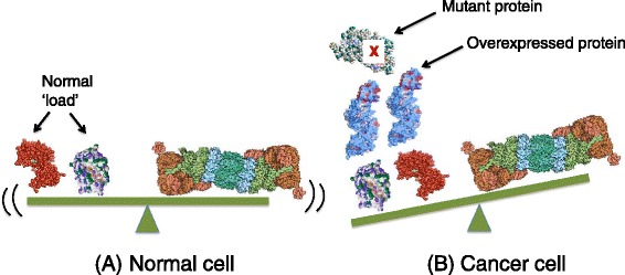
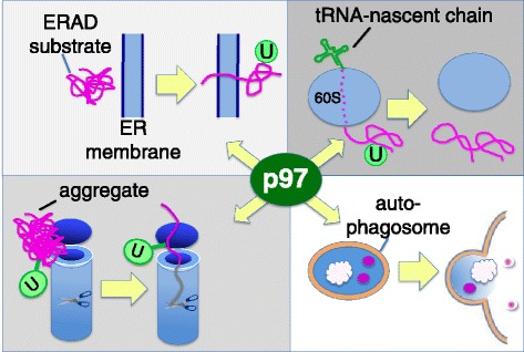
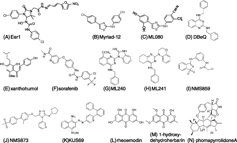
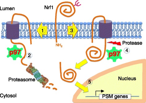
Genomic alterations may make cancer cells more dependent than normal cells on mechanisms of proteostasis, including protein folding and degradation. This proposition is the basis for the clinical use of proteasome inhibitors to treat multiple myeloma and mantle cell lymphoma. However, proteasome inhibitors have not proved effective in treating other cancers, and this has called into question the general applicability of this approach. Here, I consider possible explanations for this apparently limited applicability, and discuss whether inhibiting other broadly acting components of the ubiquitin-proteasome system - including ubiquitin-activating enzyme and the AAA-ATPase p97/VCP - might be more generally effective in cancer therapy.


Figures

Figure 1
Figure 1
Proteotoxic crisis in cancer cells. (A) In normal cells, the natural load of degradation substrates on the left is in balance with the capacity of the cellular ubiquitin-proteasome system (UPS), signified by the proteasome on the right. (B) In cancer cells, the load is increased due to expression of mutant proteins and/or expression of excess proteins due to aneuploidy. This results in an imbalance where the degradation load exceeds the capacity of the UPS.
Figure 2
Figure 2
Structure of the 26S proteasome. The 20S core of the proteasome is shown in grey. One copy of each of the β1, β2, and β5 active sites is present within each of two seven-subunit rings (the positions of which are marked with dashed blue lines) and face towards the inside of the 20S chamber. Each end of the 20S core is capped with a 19S regulatory particle, shown in various colors. The Rpt1-6 ATPase ring that abuts the 20S core is shown in blue and Rpn11 in green. Rpn10 (purple) and Rpn13 (gold) are two intrinsic ubiquitin chain-binding receptors within the 26S proteasome. The pore in the ATPase ring through which the substrate passes is indicated. Electron density within the image that corresponds to substrate is shown in red. Adapted from [19].
Figure 3
Figure 3
The sawtooth pattern of β5 inhibition in vivo and its relationship to the kinetics of cancer cell death. (A) A patient dosed with bortezomib at the beginning of day 1 experiences approximately 65% inhibition of β5 activity (shown as a red trace) in whole blood lysate. β5 activity recovers, and the patient is dosed again on day 4. (B) Zoom-in of (A) to emphasize β5’s pharmacodynamic response to a single dose. In this and the following examples, it is assumed that the kinetics of cancer cell death are a function of cell type and percentage inhibition. The example shown assumes that MM cells commit to cell death within a few hours when the proteasome is inhibited by more than 60% (>60% I), as signified by the time interval denoted by the light gray bar. On the other hand, solid tumor cells require much longer exposure (dark gray bar) to effect cell death at 60% inhibition. (C) Same as (B), except that a greater percentage inhibition of the proteasome is achieved. Even though the rate of recovery is the same as (B), it is suggested that solid tumor cells remain in the ‘kill zone’ below the dotted line sufficiently long to commit to apoptosis. Note that even though the time required for killing solid tumors (dark gray bar) is drawn the same as in (B), a greater percentage inhibition could reduce the time required to commit to apoptosis. (D) Same as (B), except the recovery curve has a shallower slope due to inhibition of new proteasome synthesis. In this hypothetical example, reducing the rate of recovery maintains proteasome inhibition in the ‘kill zone’ for a sufficiently long time to kill solid tumor cells.
Figure 4
Figure 4
Alternative strategies for testing the proteotoxic crisis hypothesis through proteasome inhibition. (A) Bortezomib (BTZ; red asterisk) has a very slow off-rate from 26S proteasome (26S; gray cylinders). Coupled with the high concentrations of proteasomes in red blood cells (RBCs), this results in sequestration of most BTZ in the RBC compartment following intravenous injection. (B) MLN9708 (purple asterisk) dissociates from proteasome six-fold faster than BTZ, enabling better equilibration throughout the body and stronger inhibition of proteasome in tumors. (C) Hypothetical pharmacodynamic response of β5 activity (red trace) in a patient repeatedly dosed (blue arrows) with an oral proteasome inhibitor during the course of a single day. Repeat dosing may suffice to keep β5 activity in the ‘kill zone’ (in this example, >60% inhibition) for a sufficiently long time interval (denoted by dark gray bar) to kill solid tumor cells. (D) Alternative drug targets in the proteasome: the Rpt1-6 ATPase, Rpn11, and the pockets in 29S outer rings that serve as docking sites for Rpt ATPases in 19S regulatory particle.
Figure 5
Figure 5
Roles of p97 in protein quality control. Upper left: p97 extracts unfolded or misassembled secretory and membrane proteins from the ER. Concomitant with extraction, substrates are conjugated with ubiquitin (green circle with U). Upper right: ribosomes that stall during translation are disassembled into 40S +60S by an upstream factor. The ubiquitin-conjugated nascent chain remains attached to tRNA and passes through the exit tunnel (dotted segment). p97 recognizes these complexes and releases the nascent chain from the ribosome. Lower left and right: protein and protein-RNA aggregates require p97 for metabolism. p97 may either disassemble aggregates so that the proteasome can degrade them (lower left), or the aggregates can be packaged into autophagosomes (lower right) for delivery to the lysosome. p97 is required for an undetermined step in autophagosome maturation.
Figure 6
Figure 6
Structures of p97 inhibitors. The inhibitors are as indicated in the figure. Inhibitors are listed in order of their first report in the literature. Myriad-19 is similar to Myriad-12 except that it lacks the chlorine atom. ML080 behaves essentially the same as the Myriad compounds. KUS69 is the most potent of a series of five structurally related compounds.
Figure 7
Figure 7
Mechanism of Nrf1 activation. Upon completion of synthesis, Nrf1 is rapidly directed into the retrotranslocation pathway (1). p97 extracts Nrf1 from the ER, and it is then fed to the proteasome (2). Because of the tight coupling between synthesis, retrotranslocation, and degradation, there is very little accumulation of Nrf1 at steady-state. In cells that are deficient in proteasome activity (3), retrotranslocation and degradation of Nrf1 become kinetically uncoupled. Accumulation of Nrf1 on the cytosolic side of the membrane renders it susceptible to cleavage by an unknown protease (5), which releases a soluble 110 kDa fragment that translocates to the nucleus and activates transcription of genes that encode proteasome subunits. PSM: proteasome components.

Similar articles

Cited by

References

    1. Hershko A, Ciechanover A. The ubiquitin system. Annu Rev Biochem. 1998;67:425–479. doi: 10.1146/annurev.biochem.67.1.425. - DOI - PubMed
    1. Balch WE, Morimoto RI, Dillin A, Kelly JW. Adapting proteostasis for disease intervention. Science. 2008;319:916–919. doi: 10.1126/science.1141448. - DOI - PubMed
    1. Vogelstein B, Papadopoulos N, Velculescu VE, Zhou S, Diaz LA, Jr, Kinzler KW. Cancer genome landscapes. Science. 2013;339:1546–1558. doi: 10.1126/science.1235122. - DOI - PMC - PubMed
    1. Weaver BA, Cleveland DW. Does aneuploidy cause cancer? Curr Opin Cell Biol. 2006;18:658–667. doi: 10.1016/j.ceb.2006.10.002. - DOI - PubMed
    1. Williams BR, Prabhu VR, Hunter KE, Glazier CM, Whittaker CA, Housman DE, Amon A. Aneuploidy affects proliferation and spontaneous immortalization in mammalian cells. Science. 2008;322:703–709. doi: 10.1126/science.1160058. - DOI - PMC - PubMed

Publication types

MeSH terms