. 2014 Nov 25;111(47):16860-5.
doi: 10.1073/pnas.1415712111. Epub 2014 Nov 10.

Surface attachment induces Pseudomonas aeruginosa virulence

Affiliations

Surface attachment induces Pseudomonas aeruginosa virulence

Albert Siryaporn et al. Proc Natl Acad Sci U S A. .

Abstract

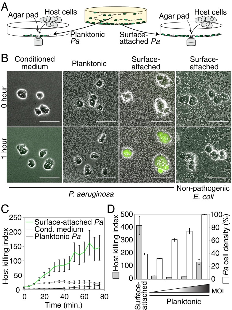
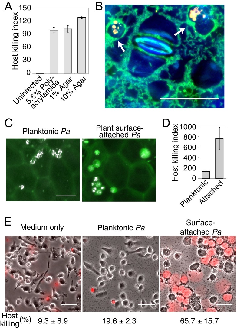
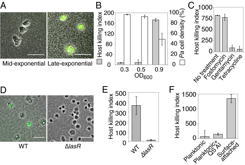
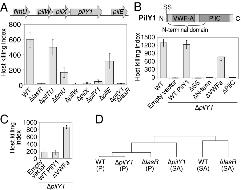
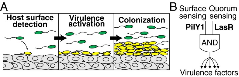
Pseudomonas aeruginosa infects every type of host that has been examined by deploying multiple virulence factors. Previous studies of virulence regulation have largely focused on chemical cues, but P. aeruginosa may also respond to mechanical cues. Using a rapid imaging-based virulence assay, we demonstrate that P. aeruginosa activates virulence in response to attachment to a range of chemically distinct surfaces, suggesting that this bacterial species responds to mechanical properties of its substrates. Surface-activated virulence requires quorum sensing, but activating quorum sensing does not induce virulence without surface attachment. The activation of virulence by surfaces also requires the surface-exposed protein PilY1, which has a domain homologous to a eukaryotic mechanosensor. Specific mutation of the putative PilY1 mechanosensory domain is sufficient to induce virulence in non-surface-attached cells, suggesting that PilY1 mediates surface mechanotransduction. Triggering virulence only when cells are both at high density and attached to a surface—two host-nonspecific cues—explains how P. aeruginosa precisely regulates virulence while maintaining broad host specificity.

Keywords: PilY1; bacterial mechanosensation; contact regulation; host detection; von Willebrand factor.


Conflict of interest statement

The authors declare no conflict of interest.

Figures

Fig. 1.
Fig. 1.
Surface attachment stimulates P. aeruginosa virulence toward D. discoideum. (A) Schematic depicting a rapid imaging-based virulence assay. Planktonic or surface-attached subpopulations of P. aeruginosa (Pa) were isolated from a Petri dish, mixed with host cells, confined to a single plane using an agar pad, and imaged. (B) Composite phase contrast (grayscale) and calcein-AM fluorescence (green) images of D. discoideum (amoebae) that were mixed with conditioned medium, planktonic or glass surface-attached P. aeruginosa, or with nonpathogenic E. coli. (Scale bars: 50 µm.) (C) Host killing indexes of amoebae that were mixed with conditioned medium or with planktonic or plastic surface-attached P. aeruginosa. (D) Host killing indexes (gray bars) and corresponding cell densities (white bars) of surface-attached or planktonic P. aeruginosa at different multiplicities of infection (MOI). Bars represent the average of three independent experiments, and error bars indicate SE.
Fig. 2.
Fig. 2.
Virulence activation is surface- and host-independent. (A) Host killing indexes of P. aeruginosa cells that were attached to polyacrylamide or agar surfaces. Error bars indicate the range of two independent experiments. (B) Assay to test virulence of P. aeruginosa (green) that attach to the surface of a plant leaf (surface and stoma in blue) using amoebae as hosts (arrows point to cell compartments labeled yellow by uptake of fluorescent beads). (C and D) Amoebae (white) were mixed with planktonic (C, Left) or plant surface-attached (C, Right) P. aeruginosa cells (not visible in image), imaged on the plant leaf surface (faint green from leaf autofluorescence), and assessed for viability by using calcein-AM (green), which was used to compute host killing indexes (D). Error bars indicate the SD of three independent experiments. (E) Composite images of phase contrast (grayscale) and propidium iodide fluorescence (red) of mouse macrophages that were exposed to medium only (Left) or to planktonic (Center) or surface-attached (Right) P. aeruginosa for 3 h. The fraction of macrophages with propidium iodide fluorescence after 4.5 h of exposure is given. (Scale bars: 50 µm.)
Fig. 3.
Fig. 3.
Contact-mediated virulence is rapidly induced and requires quorum sensing but not growth. (A and B) Composite phase contrast and calcein-AM fluorescence images (A) and host killing indexes (gray bars) and corresponding P. aeruginosa cell densities (white bars; B; see SI Appendix, SI Materials and Methods for details) for P. aeruginosa that were surface-attached for 1 h at midexponential (OD600 = 0.3 or 0.5) or late-exponential (OD600 = 0.9) growth phases and mixed with amoebae. (C) Host killing indexes for P. aeruginosa that were treated with antibiotics during surface attachment at late-exponential phase. (D and E) Composite phase contrast and calcein-AM fluorescence images (D) and host killing indexes (E) for WT and ΔlasR (quorum-sensing defective) cells. (F) Host killing indexes of planktonic P. aeruginosa cells that were supplemented with DMSO or quorum-sensing autoinducers (QS AI). Bars represent the average of at least two independent experiments, and error bars indicate SD. (Scale bars: 50 µm.)
Fig. 4.
Fig. 4.
PilY1 regulates surface-activated virulence and gene expression through a mechanosensory VWFa domain. (A) Amoebae host killing indexes of surface-attached pilY1 operon P. aeruginosa mutants. (B) Host killing indexes of surface-attached PilY1 mutants containing a deletion of the N-terminal signal sequence (SS) or deletion of the N-terminal, VWFa, or PilC domains (schematic of PilY1 domains at top). (C) Host killing indexes of planktonic (non–surface-attached) P. aeruginosa cells containing empty vector or expressing WT or VWFa-domain–deleted PilY1. All bars are the average of three independent experiments, and error bars represent SD. (D) Hierarchical clustering tree (correlation values are given in SI Appendix, Fig. S7D) for transcriptional profiles from microarrays for WT, ΔlasR, and ΔpilY1 cells for either surface-attached (SA) or planktonic (P) P. aeruginosa cells for genes that were activated by at least fourfold by surface attachment in overnight cultures.
Fig. 5.
Fig. 5.
Surface detection is a host-nonspecific signal that activates virulence. (A) Schematic of proposed model in which bacterial cells detect host surfaces through mechanical cues during initial attachment. The induction of virulence through this detection mechanism establishes an environment that is suitable for bacterial colonization of the host. (B) An ‘AND’ gate model for surface-activated virulence in which both surface sensing (mediated by PilY1) and quorum sensing (mediated by LasR) are required for activating virulence.

Comment in

References

    1. Lee DG, et al. Genomic analysis reveals that Pseudomonas aeruginosa virulence is combinatorial. Genome Biol. 2006;7(10):R90. - PMC - PubMed
    1. Feinbaum RL, et al. Genome-wide identification of Pseudomonas aeruginosa virulence-related genes using a Caenorhabditis elegans infection model. PLoS Pathog. 2012;8(7):e1002813. - PMC - PubMed
    1. Lau GW, Hassett DJ, Ran H, Kong F. The role of pyocyanin in Pseudomonas aeruginosa infection. Trends Mol Med. 2004;10(12):599–606. - PubMed
    1. Morihara K, Tsuzuki H, Oka T, Inoue H, Ebata M. Pseudomonas aeruginosa elastase. Isolation, crystallization, and preliminary characterization. J Biol Chem. 1965;240:3295–3304. - PubMed
    1. Blumer C, Haas D. Mechanism, regulation, and ecological role of bacterial cyanide biosynthesis. Arch Microbiol. 2000;173(3):170–177. - PubMed

Publication types