Review
. 2014 Oct 27:5:496.
doi: 10.3389/fimmu.2014.00496. eCollection 2014.

Eosinophil secretion of granule-derived cytokines

Affiliations
Review

Eosinophil secretion of granule-derived cytokines

Lisa A Spencer et al. Front Immunol. .

Abstract

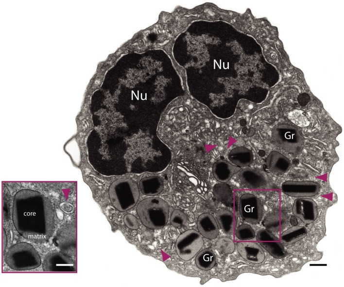
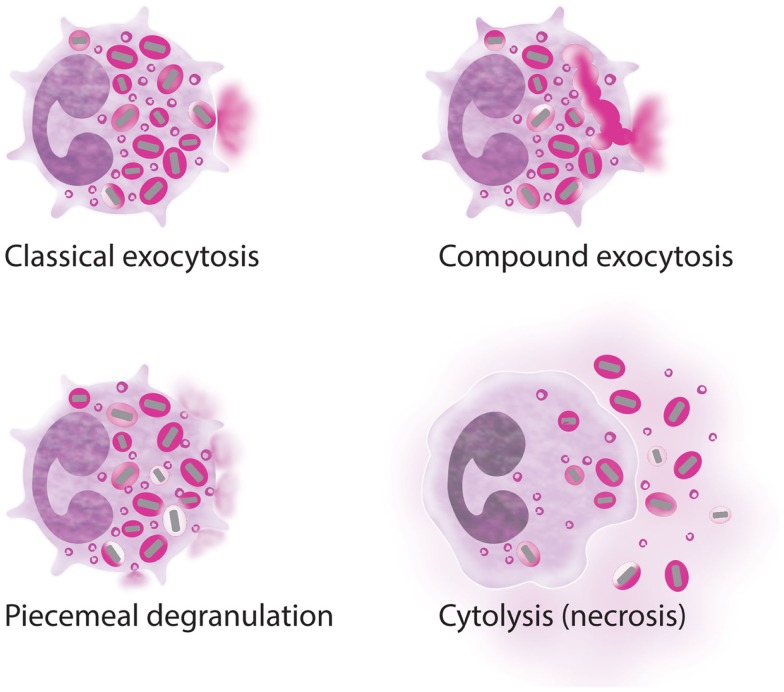
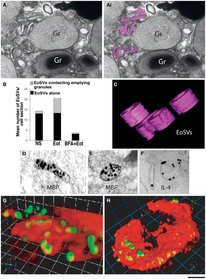
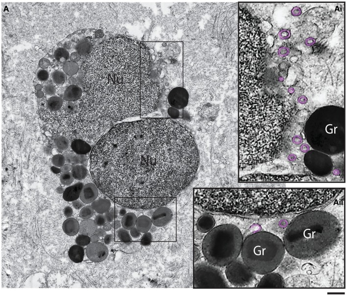
Eosinophils are tissue-dwelling leukocytes, present in the thymus, and gastrointestinal and genitourinary tracts of healthy individuals at baseline, and recruited, often in large numbers, to allergic inflammatory foci and sites of active tissue repair. The biological significance of eosinophils is vast and varied. In health, eosinophils support uterine and mammary gland development, and maintain bone marrow plasma cells and adipose tissue alternatively activated macrophages, while in response to tissue insult eosinophils function as inflammatory effector cells, and, in the wake of an inflammatory response, promote tissue regeneration, and wound healing. One common mechanism driving many of the diverse eosinophil functions is the regulated and differential secretion of a vast array of eosinophil-derived cytokines. Eosinophils are distinguished from most other leukocytes in that many, if not all, of the over three dozen eosinophil-derived cytokines are pre-synthesized and stored within intracellular granules, poised for very rapid, stimulus-induced secretion. Eosinophils engaged in cytokine secretion in situ utilize distinct pathways of cytokine release that include classical exocytosis, whereby granules themselves fuse with the plasma membrane and release their entire contents extracellularly; piecemeal degranulation, whereby granule-derived cytokines are selectively mobilized into vesicles that emerge from granules, traverse the cytoplasm and fuse with the plasma membrane to release discrete packets of cytokines; and eosinophil cytolysis, whereby intact granules are extruded from eosinophils, and deposited within tissues. In this latter scenario, extracellular granules can themselves function as stimulus-responsive secretory-competent organelles within the tissue. Here, we review the distinctive processes of differential secretion of eosinophil granule-derived cytokines.

Keywords: cytokine; cytolysis; degranulation; eosinophil; granule; piecemeal degranulation; secretion.


Figures

Figure 1
Figure 1
Transmission electron microscopy of a human eosinophil. This cell is characterized by a major population of specific granules (Gr) with a unique morphology – an internal often electron-dense crystalline core and an outer electron-lucent matrix surrounded by a delimiting trilaminar membrane. Note the typical bilobed nucleus (Nu) and large tubular carriers (arrowheads). The inset shows secretory granules and a tubular vesicle at higher magnification. Bars: 500 nm; 300 nm (inset).
Figure 2
Figure 2
Processes of eosinophil secretion. Eosinophils may secrete their granule proteins by classic exocytosis (individual granule fusion with the plasma membrane and release of the total granule content); compound exocytosis (intracellular granule–granule fusion before extracellular release); piecemeal degranulation (vesicular transport of small packets of materials from the secretory granules to the cell surface); and/or cytolysis (extracellular deposition of intact granules upon cell lysis). More than one process can be involved in inflammatory responses.
Figure 3
Figure 3
Ultrastructure of an eotaxin-activated human eosinophil showing piecemeal degranulation (PMD). (A) After stimulation, specific granules (Gr) exhibit different degrees of emptying of their contents and morphological diversity indicative of PMD, such as (B) lucent areas in their cores, (C) enlargement and reduced electron density, and (D) residual cores. Eosinophils were isolated by negative selection from healthy donors, stimulated with eotaxin-1 for 1 h, immediately fixed and prepared for transmission electron microscopy as before (43). Nu, nucleus; LB, lipid body. Scale bar: 500 nm (A); 170 nm (B–D).
Figure 4
Figure 4
Vesicular trafficking of granule-derived products from human eosinophils. (A) Eosinophil sombrero vesicles – EoSVs – [highlighted in pink in (Ai)] are observed in the cytoplasm surrounding an emptying, enlarged secretory granule (Gr). An intact granule (Gr) with typical morphology is also observed. (B) Quantification of EoSV numbers revealed significant formation of these vesicles and association with granules undergoing release of their products, after eotaxin-1 (EOT) stimulation (45). Brefeldin-A (BFA) pretreatment suppressed all EoSV numbers dramatically (P < 0.05). NS, not stimulated. (C) Three-dimensional (3D) models obtained from electron tomographic analyses show EoSVs as curved tubular and open structures surrounding a cytoplasmic center. (D–F) As demonstrated by immunonanogold electron microscopy, major basic protein (MBP) (D,E) is transported within the EoSVs lumen, while IL-4 mobilization is associated with vesicle membrane (F). In (G,H), human blood eosinophils suspended in an anti-IL-4 capture antibody-containing agarose matrix were stimulated with eotaxin-1. 3D reconstructed images demonstrate released and captured IL-4 as focal fluorescent green spots at the outer surface of the cell membrane (stained in red). (B,F–H) were reprinted from Ref. (45) and (C–E) from Ref. (46) with permission. Scale bar: 250 nm (A); 150 nm (C–F); 4 μm (G); 6 μm (H).
Figure 5
Figure 5
Ultrastructure of a tissue human eosinophil undergoing cytolysis. Note the disintegrating nucleus (Nu) and extracellular free secretory granules (Gr) in the surrounding tissue. (Ai, Aii) are boxed areas of (A) seen at higher magnification. Note the presence of free, intact eosinophil sombrero vesicles (EoSVs – highlighted in pink) in the tissue, after cell lysis. Tissue eosinophils were present in a biopsy performed on a patient with inflammatory bowel disease. Scale bar: 800 nm (A); 300 nm (Ai, Aii).

References

    1. Spry CJF. Eosinophils: A Comprehensive Review and Guide to the Scientific and Medical Literature. Oxford: Oxford Medical Publications; (1988).
    1. Acharya KR, Ackerman SJ. Eosinophil granule proteins: form and function. J Biol Chem (2014) 289(25):17406–15.10.1074/jbc.R113.546218 - DOI - PMC - PubMed
    1. Wong DT, Weller PF, Galli SJ, Elovic A, Rand TH, Gallagher GT, et al. Human eosinophils express transforming growth factor alpha. J Exp Med (1990) 172(3):673–81.10.1084/jem.172.3.673 - DOI - PMC - PubMed
    1. Moqbel R, Coughlin JJ. Differential secretion of cytokines. Sci STKE (2006) 2006(338):e26.10.1126/stke.3382006pe26 - DOI - PubMed
    1. Beil WJ, Weller PF, Tzizik DM, Galli SJ, Dvorak AM. Ultrastructural immunogold localization of tumor necrosis factor-alpha to the matrix compartment of eosinophil secondary granules in patients with idiopathic hypereosinophilic syndrome. J Histochem Cytochem (1993) 41(11):1611–5.10.1177/41.11.8409368 - DOI - PubMed

LinkOut - more resources