. 2014 Oct 29;5(4):e0032.
doi: 10.5041/RMMJ.10166. eCollection 2014 Oct.

Extracellular vesicles in hematological disorders

Affiliations

Extracellular vesicles in hematological disorders

Anat Aharon et al. Rambam Maimonides Med J. .

Abstract

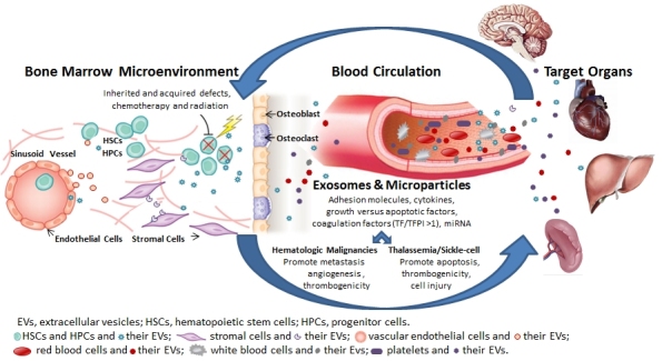
Extracellular vesicles (EVs), comprised of exosomes, microparticles, apoptotic bodies, and other microvesicles, are shed from a variety of cells upon cell activation or apoptosis. EVs promote clot formation, mediate pro-inflammatory processes, transfer proteins and miRNA to cells, and induce cell signaling that regulates cell differentiation, proliferation, migration, invasion, and apoptosis. This paper will review the contribution of EVs in hematological disorders, including hemoglobinopathies (sickle cell disease, thalassemia), paroxysmal nocturnal hemoglobinuria, and hematological malignancies (lymphomas, myelomas, and acute and chronic leukemias).

Keywords: Extracellular vesicles; exosomes; hemoglobinopathies; leukemia; lymphomas; microRNA; myeloma; thrombogenicity.


Figures

Figure 1.
Figure 1.
Involvement of Extracellular Vesicles (EVs) in Hematologic Disorders. Inherited and acquired defects as well as exposure to chemotherapy, radiation, and cytokines result in release of EVs from a variety of cells (e.g. hematopoietic stem and progenitor cells, blood, vascular, and tumor cells) to the bone marrow microenvironment, the vascular compartment, and the target organ with auto- and paracrine effects. Extracellular vesicles, which include microparticles and exosomes, express adhesion molecules, cytokines, growth versus apoptotic factors, coagulation factors and miRNA. In hematologic malignancies, EVs promote metastasis, angiogenesis, and thrombogenicity. In thalassemia/sickle cell diseases, EVs promote cell injury, apoptosis, and thrombogenicity.

References

    1. Piccin A, Murphy WG, Smith OP. Circulating microparticles: pathophysiology and clinical implications. Blood Rev. 2007;21:157–71. doi: 10.1016/j.blre.2006.09.001. - DOI - PubMed
    1. Fevrier B, Raposo G. Exosomes: endosomal-derived vesicles shipping extracellular messages. Curr Opin Cell Biol. 2004;16:415–21. doi: 10.1016/j.ceb.2004.06.003. - DOI - PubMed
    1. Bobrie A, Colombo M, Krumeich S, et al. Diverse subpopulations of vesicles secreted by different intracellular mechanisms are present in exosome preparations obtained by differential ultracentrifugation. J Extracell Vesicles. 2012;1:18397. doi: 10.3402/jev.v1i0.18397. - DOI - PMC - PubMed
    1. Colombo M, Moita C, van Niel G, et al. Analysis of ESCRT functions in exosome biogenesis, composition and secretion highlights the heterogeneity of extracellular vesicles. J Cell Sci. 2013;126:5553–65. doi: 10.1242/jcs.128868. - DOI - PubMed
    1. Gan X, Gould SJ. Identification of an inhibitory budding signal that blocks the release of HIV particles and exosome/microvesicle proteins. Mol Biol Cell. 2011;22:817–30. doi: 10.1091/mbc.E10-07-0625. - DOI - PMC - PubMed

LinkOut - more resources