. 2014 Oct 11:4:56.
doi: 10.1186/s13550-014-0056-0. eCollection 2014.

U-SPECT-BioFluo: an integrated radionuclide, bioluminescence, and fluorescence imaging platform

Affiliations

U-SPECT-BioFluo: an integrated radionuclide, bioluminescence, and fluorescence imaging platform

Matthias N van Oosterom et al. EJNMMI Res. .

Abstract

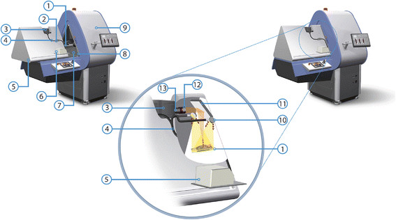
Background: In vivo bioluminescence, fluorescence, and single-photon emission computed tomography (SPECT) imaging provide complementary information about biological processes. However, to date these signatures are evaluated separately on individual preclinical systems. In this paper, we introduce a fully integrated bioluminescence-fluorescence-SPECT platform. Next to an optimization in logistics and image fusion, this integration can help improve understanding of the optical imaging (OI) results.

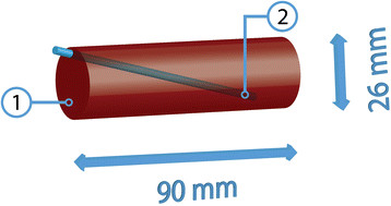
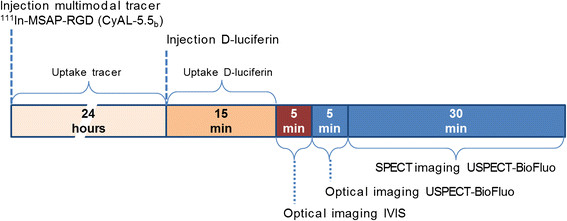
Methods: An OI module was developed for a preclinical SPECT system (U-SPECT, MILabs, Utrecht, the Netherlands). The applicability of the module for bioluminescence and fluorescence imaging was evaluated in both a phantom and in an in vivo setting using mice implanted with a 4 T1-luc + tumor. A combination of a fluorescent dye and radioactive moiety was used to directly relate the optical images of the module to the SPECT findings. Bioluminescence imaging (BLI) was compared to the localization of the fluorescence signal in the tumors.

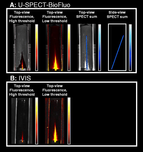
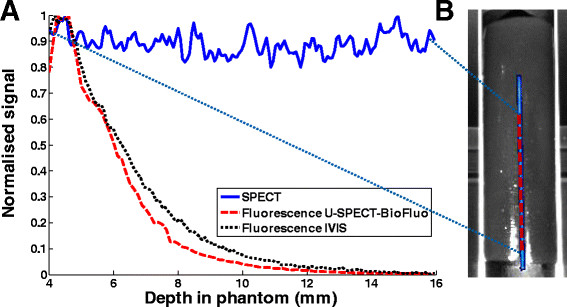
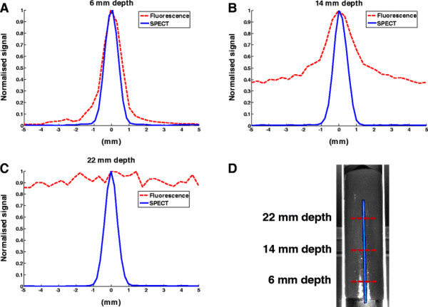
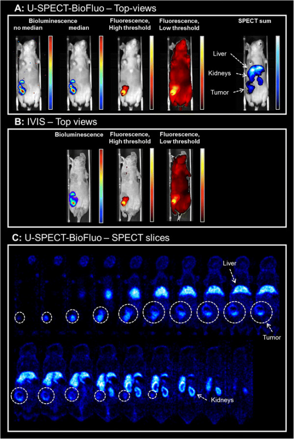
Results: Both the phantom and in vivo mouse studies showed that superficial fluorescence signals could be imaged accurately. The SPECT and bioluminescence images could be used to place the fluorescence findings in perspective, e.g. by showing tracer accumulation in non-target organs such as the liver and kidneys (SPECT) and giving a semi-quantitative read-out for tumor spread (bioluminescence).

Conclusions: We developed a fully integrated multimodal platform that provides complementary registered imaging of bioluminescent, fluorescent, and SPECT signatures in a single scanning session with a single dose of anesthesia. In our view, integration of these modalities helps to improve data interpretation of optical findings in relation to radionuclide images.

Keywords: Bioluminescence imaging; Fluorescence imaging; Multimodal molecular imaging; Nuclear medicine; SPECT; Small animal.


Figures

Figure 1
Figure 1
Overview of the U-SPECT-BioFluo platform. (1) Animal bed. (2) Excitation filter box. (3) Camera. (4) Optical fiber bundle. (5) Dark box. (6) Light source. (7) Black ABS-plastic insert. (8) U-profile light lock. (9) U-SPECT. (10) Mirrors for excitation light. (11) Mirror directing light from the bed to the camera. (12) Lens hood and emission filter holder. (13) Camera lens.
Figure 2
Figure 2
Schematic diagram of the tissue simulating gel phantom. (1) Phantom. (2) Fluorescent and radioactive capillary.
Figure 3
Figure 3
Protocol timeline for in vivo measurements. The injection moments of 111In-MSAP-RGD (CyAL-5.5b dye), D-Luciferin, and subsequent imaging periods are indicated.
Figure 4
Figure 4
Phantom imaging results. (A) Fluorescence (500 ms exposure time) and SPECT imaging on the U-SPECT-BioFluo. (B) Fluorescence imaging on the IVIS (1s exposure time).
Figure 5
Figure 5
Effects of depth-dependent attenuation. (A) Fluorescence signal (phantom background level subtracted) and SPECT signal of capillary versus depth in gel phantom. The maximum SPECT and fluorescence signals have been normalized to 1. (B) The positions of the axial line profiles are indicated in the top-view image of the phantom.
Figure 6
Figure 6
Comparison of image blurring for SPECT and fluorescence. (A,B,C) SPECT and fluorescence line profiles orthogonal to the capillary at different depths in the phantom. The maximum signal of each profile has been normalized to 1. (D) The positions of the line profiles are indicated in the top-view image.
Figure 7
Figure 7
Imaging results for a mouse with a 4 T1-luc + tumor. Bioluminescence images are obtained by D-Luciferin injection. Fluorescence and SPECT images are obtained by injection of multimodal tracer 111In-RGD-MSAP. (A) U-SPECT-BioFluo. Bioluminescent images both with and without median filter applied. (B) IVIS. (C) U-SPECT-BioFluo: 1-mm thick horizontal SPECT slices (anterior to posterior). Organs have been indicated in the SPECT images. White circles indicate the tumor location in the slices.

References

    1. Weissleder R, Mahmood U. Molecular imaging. Radiology. 2001;219:316–333. doi: 10.1148/radiology.219.2.r01ma19316. - DOI - PubMed
    1. Ramaswamy AK, Hamilton M, Joshi RV, Kline BP, Li R, Wang P, Goergen CJ. Molecular imaging of experimental abdominal aortic aneurysms. Scientific World Journal. 2013;2013:973150. doi: 10.1155/2013/973150. - DOI - PMC - PubMed
    1. Culver J, Akers W, Achilefu S. Multimodality molecular imaging with combined optical and SPECT/PET modalities. J Nucl Med. 2008;49:169–172. doi: 10.2967/jnumed.107.043331. - DOI - PubMed
    1. Deroose CM, De A, Loening AM, Chow PL, Ray P, Chatziioannou AF, Gambhir SS. Multimodality imaging of tumor xenografts and metastases in mice with combined small-animal PET, small-animal CT, and bioluminescence imaging. J Nucl Med. 2007;48:295–303. - PMC - PubMed
    1. Park JM, Gambhir SS. Multimodality radionuclide, fluorescence, and bioluminescence small-animal imaging. Proc IEEE. 2005;93:771–783. doi: 10.1109/JPROC.2005.844263. - DOI