. 2015 Mar 15;24(6):768-80.
doi: 10.1089/scd.2014.0225. Epub 2014 Dec 22.

Label-retaining stromal cells in mouse endometrium awaken for expansion and repair after parturition

Affiliations

Label-retaining stromal cells in mouse endometrium awaken for expansion and repair after parturition

Mingzhu Cao et al. Stem Cells Dev. .

Abstract

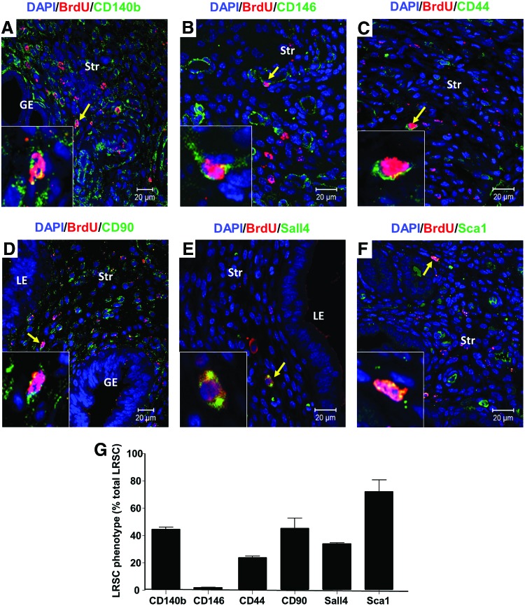
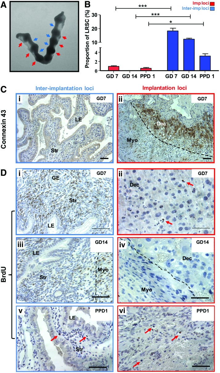
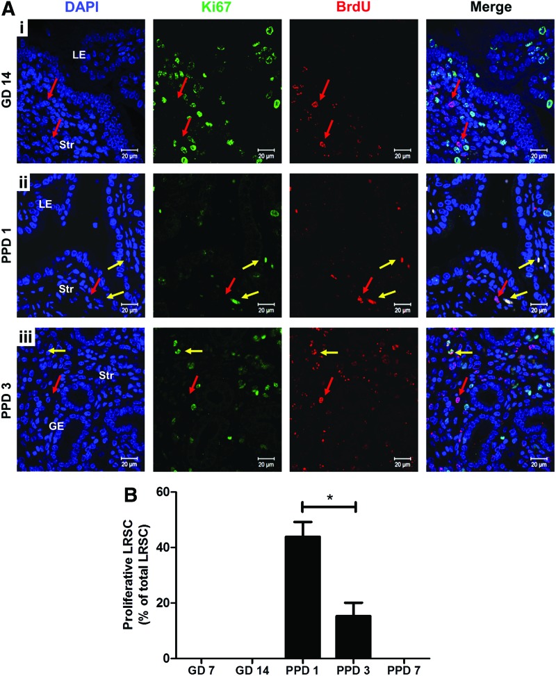
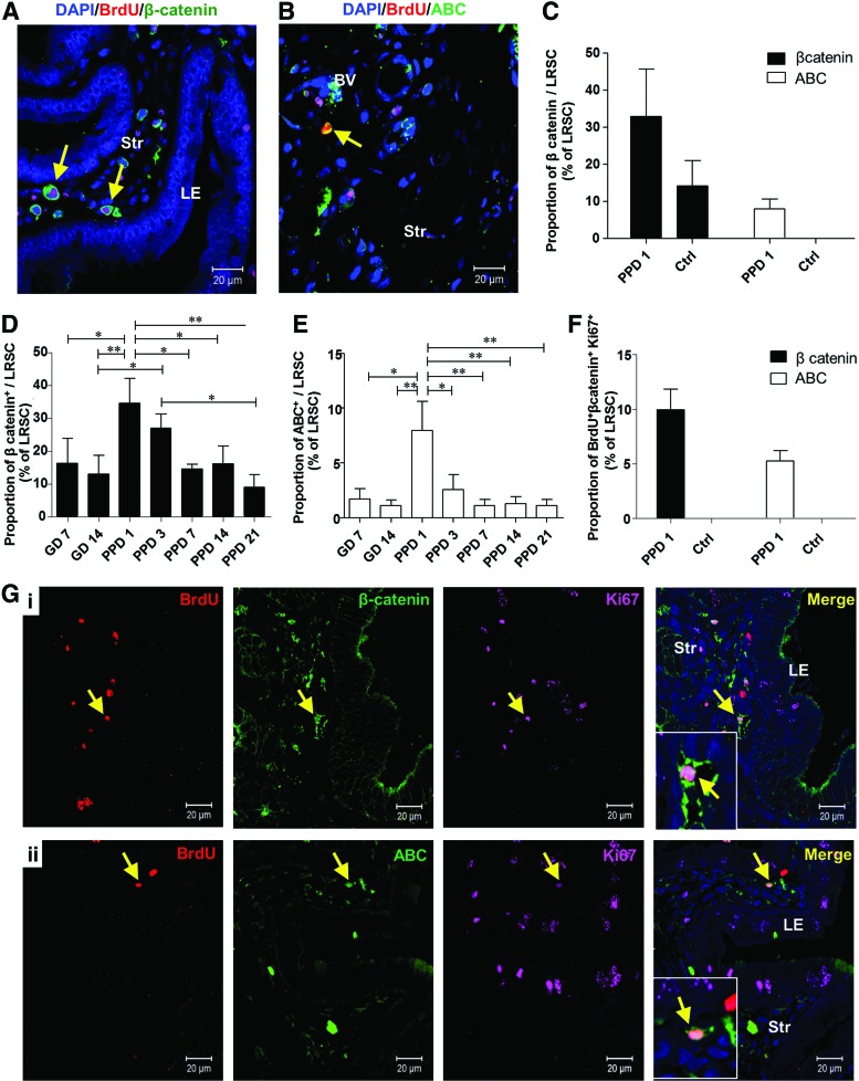
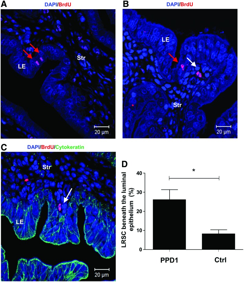
Human and mouse endometrium undergo dramatic cellular reorganization during pregnancy and postpartum. Somatic stem cells maintain homeostasis of the tissue by providing a cell reservoir for regeneration. We hypothesized that endometrial cells with quiescent properties (stem/progenitor cells) were involved in the regeneration of the endometrial tissue. Given that stem cells divide infrequently, they can retain the DNA synthesis label [bromodeoxyuridine (BrdU)] after a prolonged chase period. In this study, prepubertal mice were pulsed with BrdU and after a 6-week chase a small population of label-retaining stromal cells (LRSC) was located primarily beneath the luminal epithelium, adjacent to blood vessels, and near the endometrial-myometrial junction. Marker analyses suggested that they were of mesenchymal origin expressing CD44(+), CD90(+), CD140b(+), CD146(+), and Sca-1(+). During pregnancy, nonproliferating LRSC predominately resided at the interimplantation/placental loci of the gestational endometrium. Immediately after parturition, a significant portion of the LRSC underwent proliferation (BrdU(+)/Ki-67(+)) and expressed total and active β-catenin. The β-catenin expression in the LRSC was transiently elevated at postpartum day (PPD) 1. The proliferation of LRSC resulted in a significant decline in the proportion of LRSC in the postpartum uterus. The LRSC returned to dormancy at PPD7, and the percentage of LRSC remained stable thereafter until 11 weeks. This study demonstrated that LRSC can respond efficiently to physiological stimuli upon initiation of uterine involution and return to its quiescent state after postpartum repair.


Figures

<b>FIG. 1.</b>
FIG. 1.
Quantitative analysis of BrdU-labeled cells in gestational and postpartum endometrium. (A) Timeline of the experimental methods. Prepubertal (day 19) C57BL/6J female mice were pulse labeled with BrdU (50 μg/g of body weight) twice daily for 4 days, and the label was then chased for up to 11 weeks. Female mice at 6 weeks of age were naturally mated with fertile males. Pregnant female mice were identified by vaginal plugs. Tissue was collected 4 h after initial labeling (as denoted by formula image
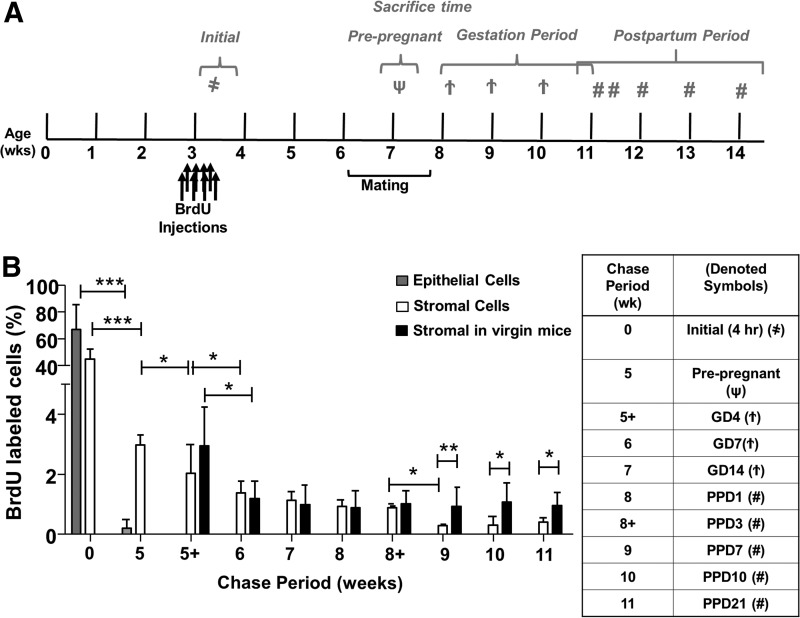
), prepregnancy (as denoted by ψ), GD: 4, 7, 14 (as denoted by formula image), and PPD: 1, 3, 7, 14, 21 (as denoted by #). (B) BrdU-labeled cells are expressed as a percentage of total epithelial or stromal cells. Data are expressed as mean±SEM, n=3–5 per group. BrdU, bromodeoxyuridine; GD, gestational days; PPD, postpartum days; SEM, standard error of the mean. *P<0.05; **P<0.01; ***P<0.001.
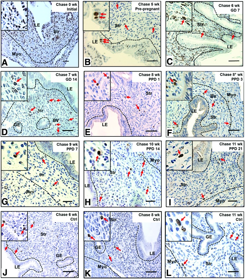
<b>FIG. 2.</b>
FIG. 2.
Localization of label-retaining cells in (A) prepubertal, (B) prepregnant, (C, D) GD, (EI) PPD, and (JL) virgin mouse endometrium. (A) BrdU immunohistochemistry of prepubertal-labeled mouse uteri 4 h after the last injection (initial labeling, 0-week chase) showed majority of epithelial and stromal cells labeled with BrdU. (B) Before pregnancy, a few epithelial cells still retain BrdU labeling, but stromal BrdU-labeled cells rapidly declined. (C, D) During gestation, no epithelial BrdU-labeled cells were observed, and stromal BrdU-labeled cells were enriched in the nonimplantation/placental locus. (EI) From PPD3 to PPD21, several LRSC localized in the vicinity of blood vessels and adjacent to the luminal epithelium or endometrial–myometrium junction. (JL) Stromal BrdU-labeled cells were detected in virgin mice at similar locations as remodeling mice. Inserts are enlarged figures of BrdU-labeled cells at different time points. Red arrows show the distribution of BrdU-labeled cells. LE, luminal epithelium; GE, glandular epithelium; Str, stroma; Myo, myometrium; BV, blood vessel; LRSC, label-retaining stromal cells. Scale bar: 5 μm. Color images available online at www.liebertpub.com/scd
<b>FIG. 3.</b>
FIG. 3.
Phenotypic characterization of LRSC with various surface markers. Immunofluorescence images showing the LRSC and their colocalization (yellow arrow) with (A) CD140b, (B) CD146, (C) CD44, (D) CD90, (E) Sall4, and (F) Sca-1 at PPD21. Inserts are enlarged figures of LRSC coexpressing different phenotypic markers. (G) The percentage of LRSC coexpressing various surface markers. Data are expressed as mean±SEM, n=3 per group. Scale bar: 20 μm. Color images available online at www.liebertpub.com/scd
<b>FIG. 4.</b>
FIG. 4.
Spatial distribution of LRSC within the pregnant and early postpartum endometrium. (A) Mouse uterine horns at GD7 showing the implantation/placental loci (red arrowheads) and interimplantation/placental loci (blue arrowheads). (B) The percentage of LRSC in interimplantation/placental loci and implantation/placental loci at different gestational and postpartum time points. (C) Immunohistochemical staining of connexin 43 (brown) localizing in the (ii) implantation/placental loci of GD7 endometrium compared with the (i) interimplantation/placental loci. (D) Representative BrdU immunohistochemistry staining (brown, red arrows) showing LRSC enriched in interimplantation/placental loci (left panel) of (i) GD7, (iii) 14, and (v) PPD1 mouse uterus. Fewer LRSC were found within implantation/placental loci (right panel) of (ii) GD7, (iv) 14, and (vi) PPD1 mouse endometrium. Data are expressed as mean±SEM, n=3 per group. Dec, decidua; Imp loci, implantation/placental loci; InterImp loci, interimplantation/placental loci. *P<0.05, ***P<0.001. Scale bar: 5 μm. Color images available online at www.liebertpub.com/scd
<b>FIG. 5.</b>
FIG. 5.
Proliferation of LRSC. (A) Representative immunofluorescent images showing LRSC colocalizing with proliferating marker Ki67 (yellow arrow) at (ii) PPD1 and (iii) PPD3. No colocalization with Ki67 (red arrow) for LRSC at GD7 and (i) GD14 uterus. (B) The percentage of proliferating LRSC at different gestational and postpartum time points. Data are expressed as mean±SEM, n=3–5 per group. *P<0.05. Scale bar: 20 μm. Color images available online at www.liebertpub.com/scd
<b>FIG. 6.</b>
FIG. 6.
Proliferating LRSC express total β-catenin and ABC. Representative immunofluorescence images show LRSC at PPD1 colocalizing with (A) β-catenin (yellow arrow) and (B) ABC (yellow arrow). (C) The percentage of LRSC coexpressing β-catenin and ABC in PPD1 and virgin mice. The percentage of LRSC coexpressing β-catenin (D) and ABC (E) at different gestational and postpartum time points. (F) The colocalization of LRSC with both β-catenin/Ki67 and ABC/Ki67 in PPD1 and virgin mice. (G) Representative figures show the triple staining of either (i) BrdU/β-catenin/Ki67 (yellow arrow) or (ii) BrdU/ABC/Ki67 (yellow arrow). Inserts are enlarged figures of LRSC coexpressing two proteins. Data are expressed as mean±SEM, n=3–5 per group. ABC, active β-catenin; Ctrl, virgin mice. *P<0.05, **P<0.01. Scale bar: 20 μm. Color images available online at www.liebertpub.com/scd
<b>FIG. 7.</b>
FIG. 7.
Identification of epithelial BrdU+ cells and location of LRSC on PPD1. (A) Epithelial BrdU+ cells in the luminal epithelium (red arrow) and (B) LRSC (white arrow) were detected within 6 h after parturition. (C) PPD1 endometrium was double immunostained with BrdU (red) and cytokeratin (green) to visualize LRSC in proximity to the luminal epithelium (white arrow). (D) Percentage of LRSC beneath the luminal epithelium (within one-cell diameter). Data are expressed as mean±SEM, n=3 per group. *P<0.05. Scale bar: 20 μm. Color images available online at www.liebertpub.com/scd

References

    1. Gargett CE. and Masuda H. (2010). Adult stem cells in the endometrium. Mol Hum Reprod 16:818–834 - PubMed
    1. Wood GA, Fata JE, Watson KL. and Khokha R. (2007). Circulating hormones and estrous stage predict cellular and stromal remodeling in murine uterus. Reproduction 133:1035–1044 - PubMed
    1. Gargett CE, Chan RW. and Schwab KE. (2007). Endometrial stem cells. Curr Opin Obstet Gynecol 19:377–383 - PubMed
    1. Ramathal CY, Bagchi IC, Taylor RN. and Bagchi MK. (2010). Endometrial decidualization: of mice and men. Semin Reprod Med 28:17–26 - PMC - PubMed
    1. Salamonsen LA. (2003). Tissue injury and repair in the female human reproductive tract. Reproduction 125:301–311 - PubMed

Publication types

LinkOut - more resources