. 2014 Nov 11;9(11):e112115.
doi: 10.1371/journal.pone.0112115. eCollection 2014.

Advanced glycation endproducts interfere with adhesion and neurite outgrowth

Affiliations

Advanced glycation endproducts interfere with adhesion and neurite outgrowth

Dorit Bennmann et al. PLoS One. .

Abstract

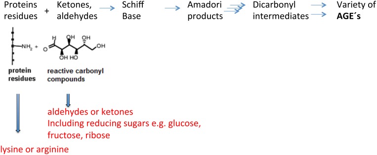
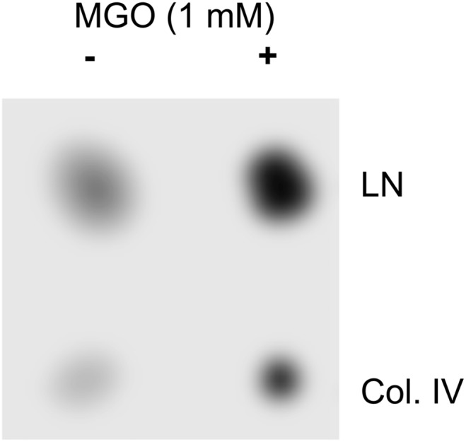
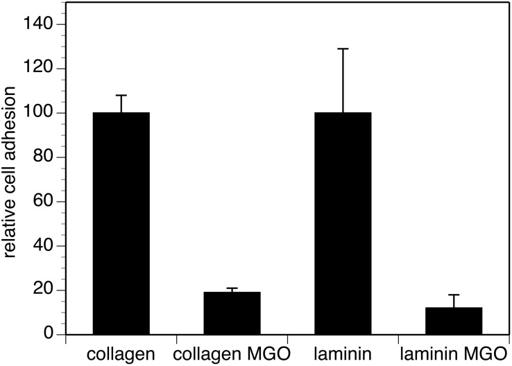
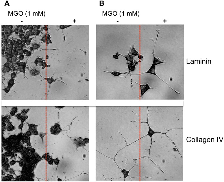
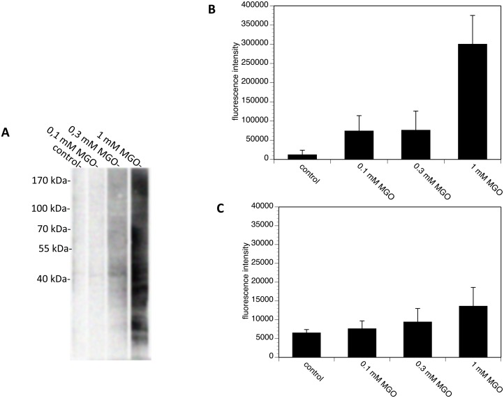
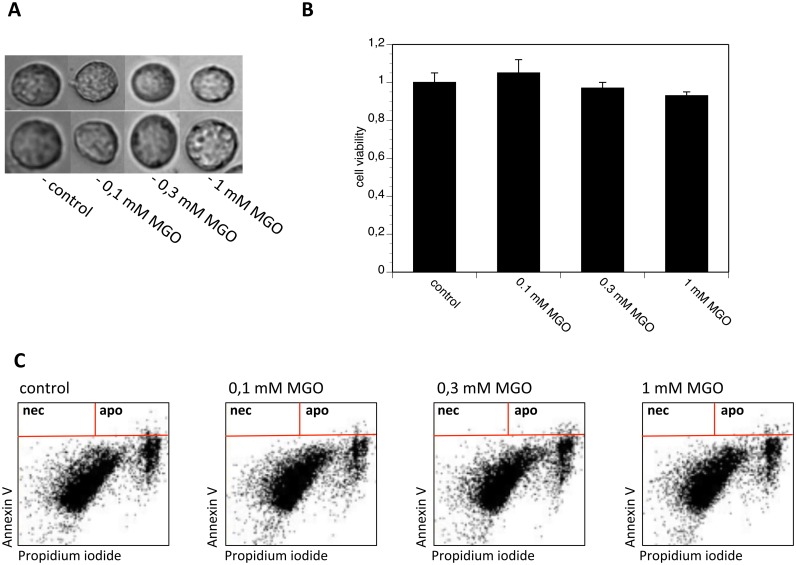
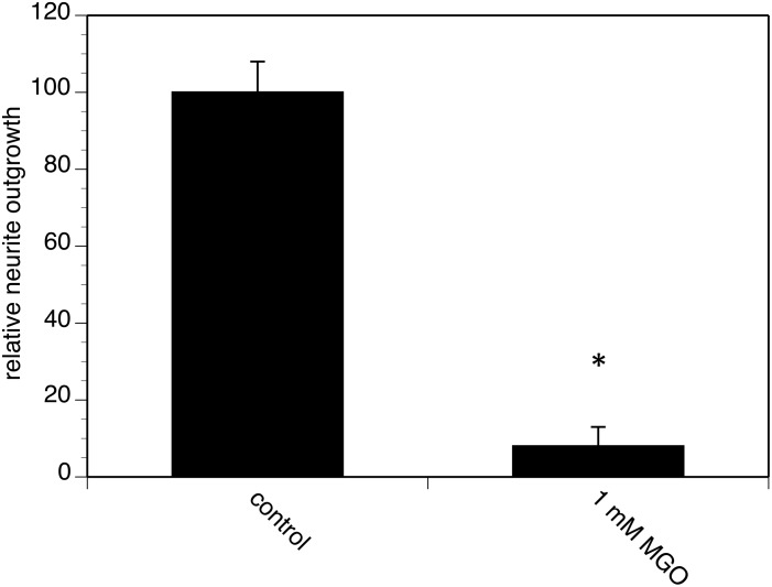
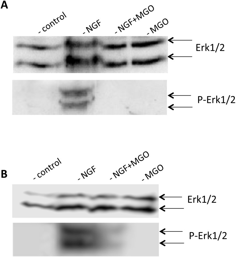
Advanced glycation endproducts (AGEs) represent a non-enzymatic posttranslational protein modification. AGEs are generated by a series of chemical reactions of free reducing monosaccharides, such as glucose, fructose or metabolites of the monosaccharide metabolism with amino groups of proteins. After oxidation, dehydration and condensation, stable AGE-modifications are formed. AGE-modified proteins accumulate in all cells and tissues as a normal feature of ageing and correlate with the glucose concentration in the blood. AGEs are increased in diabetic patients and play a significant role in the pathogenesis of most age-related neural disorders, such as Alzheimer's disease. We examined the role of AGEs on neurite outgrowth of PC12 cells. We induced the formation of AGEs using the reactive carbonyl compound methylglyoxal (MGO) as a physiological metabolite of glucose. We found that AGE-modification of laminin or collagen interfered with adhesion but not with neurite outgrowth of PC12 cells. Furthermore, the AGE-modification of PC12 cell proteins reduced NGF-induced neurite outgrowth. In conclusion, our data show that AGEs negatively influence neural plasticity.


Conflict of interest statement

Competing Interests: The authors have declared that no competing interests exist.

Figures

Figure 1
Figure 1. Formation of advanced glycation endproducts (AGE).
Figure 2
Figure 2. AGE modification of ECM proteins using MGO.
20 µg collagen IV (Col. IV) or laminin (LN) were spotted on a nitrocellulose membrane. One half of the membrane was incubated with 1 mM MGO for 4 hours. AGE formation was detected by dot blot analysis using the monoclonal CML26 antibody.
Figure 3
Figure 3. Cell adhesion of PC12 cells to AGE-modified ECM proteins.
AGE-modified and non-modified collagen IV and laminin (shown in Fig. 2) were coated on E-plates. Cell adhesion of 5×10⁵ PC12 cells was quantified by RTCA real time analysis as described. Adhesion to non-modified ( = control) substrates was set to 100% and adhesion to AGE-modified substrates was calculated in % of control. Each bar represents values of three independent experiments carried out in triplicates (*p≤0.0001).
Figure 4
Figure 4. Cell adhesion and neurite outgrowth at the border of AGE-modified ECM proteins.
A. Representative micrographs of PC12 cells taken at the border (red line) of AGE-modified or non-AGE-modified laminin or collagen IV coated cell culture plates. Three independent experiments were performed. Note that PC12 cells prefer non-AGE-modified laminin or collagen IV. B. Representative micrographs of neurite outgrowth at the border of AGE-modified or non-AGE-modified laminin or collagen IV coated cell culture plates. Three independent experiments were performed. Note that neurite outgrowth of PC12 cells was not different between non-AGE-modified laminin or collagen IV or AGE-modified laminin and collagen IV.
Figure 5
Figure 5. AGE-modification of PC12 cells using MGO.
PC12 cells were incubated with PBS, 0.1 mM MGO, 0.3 mM MGO or 1.0 mM MGO for 4 hours. A. Washed cells were solubilized and subjected to SDS-gel electrophoresis. Proteins were blotted and detected using monoclonal CML26 antibody B&C. Permeabilized (B) and non-permeabilized (C) were analyzed by flow cytometry using monoclonal CML26 antibody.
Figure 6
Figure 6. Cell viability after MGO treatment.
PC12 cells were incubated with PBS, 0.1 mM MGO, 0.3 mM MGO or 1 mM MGO for 4 hours. A. Micrographs of typical PC12 cells. B. Tryphan blue staining of PC12 cells. Bars represent three independent experiments carried out in quadruplicates. Cell viability of cells cultured in the presence of PBS ( = control) was set to 1 and cell viability expressed in relation to the control. C. FACS analysis of PC12 cells stained with annexin V and propidium iodide.
Figure 7
Figure 7. Real-time analysis of neurite outgrowth of AGE-modified PC12 cells.
PC12 cells were AGE-modified using 1 mM MGO for 4 h as shown in Figs. 4 &5. Cells were cultured on LN-coated E-plates and neurite outgrowth was induced by application of 100 ng/ml NGF. Neurite outgrowth was continuously quantified over 48 hours by RTCA as described in Pollscheit et al. (2012). Total neurite outgrowth of non-modified control cells during 48 h was set to 100% and neurite outgrowth of AGE-modified cells was expressed in % of control. Bars represent two independent experiments carried out in quadruplicates.
Figure 8
Figure 8. ERK1/2 activation of AGE-modified PC12 cells.
PC12 cells were incubated with PBS or 1 mM MGO for 4 hours and stimulated with 100 ng/ml NGF. A. Cells were harvested, washed, and solubilized. Solubilizates were centrifuged and subjected to SDS-gel electrophoresis. Proteins were blotted and detected using monoclonal ERK1/2 antibody for equal loading and monoclonal phospho ERK1/2 antibody for the detection of ERK activation. B. After MGO-treatment, cells were cultured for further 72 hours, harvested, washed, and solubilized. Solubilizates were centrifuged and subjected to SDS-gel electrophoresis. Proteins were blotted and detected using monoclonal ERK1/2 antibody for equal loading and monoclonal phospho-ERK1/2 antibody for the detection of ERK activation.

References

    1. Ansari NA, Moinuddin, Ali R (2011) Glycated lysine residues: a marker for non-enzymatic protein glycation in age-related diseases. Dis Markers 30: 317–324. - PMC - PubMed
    1. Ott C, Jacobs K, Haucke E, Navarrete Santos A, et al. (2014) Role of advanced glycation end products in cellular signaling. Redox Biol 2: 411–429. - PMC - PubMed
    1. Singh R, Barden A, Mori T, Beilin L (2001) Advanced glycation end-products: A review. Diabetologia 44: 129–146. - PubMed
    1. Requena V, Villena A, Díaz F, González F, Ríus F, et al. (1996) The effect of ageing on neurons in the visual sector of the thalamic reticular nucleus. Mech Ageing Dev 89: 185–193. - PubMed
    1. Krautwald M, Münch G (2010) Advanced glycation end products as biomarkers and gerontotoxins - A basis to explore methylglyoxal-lowering agents for Alzheimer’s disease?. Exp Gerontol 45: 744–751. - PubMed

Publication types

LinkOut - more resources