Review
. 2015:77:379-406.
doi: 10.1146/annurev-physiol-021014-071931. Epub 2014 Oct 29.

Cilia dysfunction in lung disease

Affiliations
Review

Cilia dysfunction in lung disease

Ann E Tilley et al. Annu Rev Physiol. 2015.

Abstract

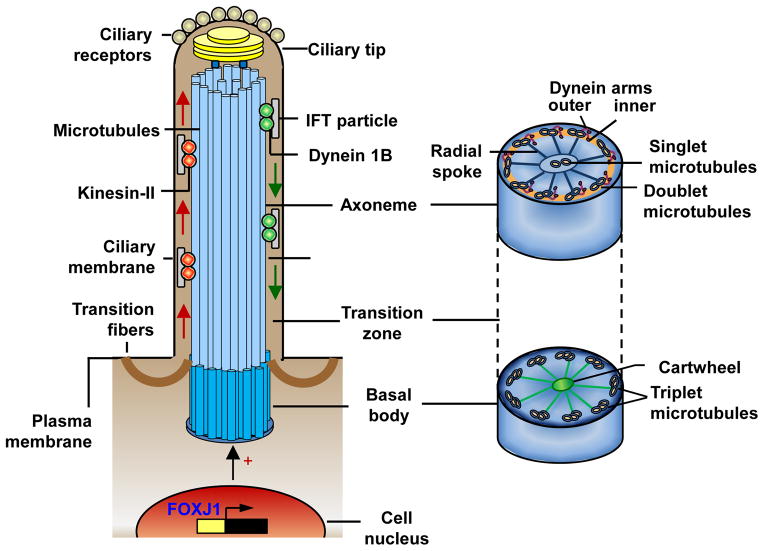
A characteristic feature of the human airway epithelium is the presence of ciliated cells bearing motile cilia, specialized cell surface projections containing axonemes composed of microtubules and dynein arms, which provide ATP-driven motility. In the airways, cilia function in concert with airway mucus to mediate the critical function of mucociliary clearance, cleansing the airways of inhaled particles and pathogens. The prototypical disorder of respiratory cilia is primary ciliary dyskinesia, an inherited disorder that leads to impaired mucociliary clearance, to repeated chest infections, and to the progressive destruction of lung architecture. Numerous acquired lung diseases are also marked by abnormalities in both cilia structure and function. In this review we summarize current knowledge regarding airway ciliated cells and cilia, how they function to maintain a healthy epithelium, and how disorders of cilia structure and function contribute to inherited and acquired lung disease.

Keywords: airway; cilia; epithelium; mucociliary escalator.


Figures

Figure 1
Figure 1
The human airway epithelium. A. Histology of the large airway epithelium (4th–5th generation of bronchi) from a healthy nonsmoker. Shown are ciliated cells, secretory cells, intermediate undifferentiated cells, and basal cells. Hematoxylin and eosin, scale bar = 20 μm. B. Ciliated cells isolated from the human small airway epithelium (10th–12th generation of bronchi) obtained by bronchoscopic brushings from a normal healthy individual. Diff-Quik, scale bar = 10 μm. C. Role of cilia in airway mucociliary clearance. The mucociliary escalator is composed of the mucus gel layer, periciliary layer, and ciliated cells. The gel-forming mucins produced by mucous secretory (“goblet”) cells are major constituents of the mucus gel layer, which entraps microorganisms and other inhaled particles and transports them out of the lung through cilia beating. The membrane-bound mucins form a brush-like pericellular niche around the cilia that controls the distribution of water between the 2 layers. During the power forward stroke, the ciliary tips extend upward into the mucus gel layer, propelling the mucus forward. During the slow return stroke, the cilia recede and are contained completely in the periciliary layer. Normal cilia length (4 to 7 μm, depending on the airway region) is critical for effective mucociliary clearance.
Figure 2
Figure 2
Ciliated cell differentiation in the human airway epithelium. A. Differentiation pathways. The basal stem/progenitor cells are capable of self-renewal and are responsible for generation of intermediate progenies, which, upon distinct activation programs, differentiate into ciliated or secretory cells. Ciliated cell differentiation depends on transcription factor forkhead box J1 (FOXJ1), whereas generation of secretory cells requires activation of Notch signaling and/or SAM pointed domain containing ETS transcription factor (SPDEF). B. Differentiation of human airway basal cells (day 0) into ciliated airway epithelium (day 28) in air-liquid interface culture. Appearance of ciliated cells is demonstrated by expression of β-tubulin IV (red signal, immunofluorescence). Cell nuclei stained with 4′,6-diamidino-2-phenylindole (DAPI; blue signal). Scale bar = 10 μm.
Figure 3
Figure 3
Structure and maintenance of human airway cilia. Each airway cilium is comprised of a “9+2” axoneme with 9 doublet microtubules and a central pair of microtubule singlets, surrounded by a specialized ciliary membrane. The ciliary membrane is contiguous with, but distinct from, the plasma membrane and harbors a number of receptors, critical for sensing environmental signals. Cilia formation is initiated and coordinated by a distinct gene expression program, primarily by activation of the transcription factor forkhead box J1 (FOXJ1). Cilia assembly begins with formation of the basal body from the centrosome, which migrates to and docks on the cell surface. The basal body is a specialized centriole with a “9+3” microtubule structure and a cartwheel embedded in pericentriolar material and anchored to the plasma membrane by transition fibers. Axonemal microtubule doublets arise from the inner 2 microtubules of the basal body microtubule triplets, extend from the basal body and form the ciliary membrane by pushing out an extension of the plasma membrane. Axonemal microtubules are elongated distally via in-traflagellar transport (IFT) of proteins, which are synthesized in the cell and moved as IFT particles by kinesin-2 motors from the basal body to the ciliary tip (anterograde IFT, left side) and by the cytoplasmic dynein motors back to the basal body (retrograde IFT, right side). The ciliary tip contains the microtubule “+” ends, from which the axonemes grow, and a number of signaling components responsible for the sensory function of cilia. The force needed for cilia beating is produced by the outer and inner dynein arms of the axonemal microtubule doublets connected to the central pair of microtubules by radial spokes.

References

    1. Mercer RR, Russell ML, Roggli VL, Crapo JD. Cell number and distribution in human and rat airways. Am J Respir Cell Mol Biol. 1994;10:613–24. - PubMed
    1. Crystal RG, Randell SH, Engelhardt JF, Voynow J, Sunday ME. Airway epithelial cells: current concepts and challenges. Proc Am Thorac Soc. 2008;5:772–7. - PMC - PubMed
    1. Knight DA, Holgate ST. The airway epithelium: structural and functional properties in health and disease. Respirology. 2003;8:432–46. - PubMed
    1. Knowles MR, Boucher RC. Mucus clearance as a primary innate defense mechanism for mammalian airways. J Clin Invest. 2002;109:571–7. - PMC - PubMed
    1. Raman T, O’Connor TP, Hackett NR, Wang W, Harvey BG, et al. Quality control in microarray assessment of gene expression in human airway epithelium. BMC Genomics. 2009;10:493. - PMC - PubMed

Publication types