Review
. 2015:77:229-49.
doi: 10.1146/annurev-physiol-021014-071727. Epub 2014 Oct 24.

Regeneration and repair of the exocrine pancreas

Affiliations
Review

Regeneration and repair of the exocrine pancreas

L Charles Murtaugh et al. Annu Rev Physiol. 2015.

Abstract

Pancreatitis is caused by inflammatory injury to the exocrine pancreas, from which both humans and animal models appear to recover via regeneration of digestive enzyme-producing acinar cells. This regenerative process involves transient phases of inflammation, metaplasia, and redifferentiation, driven by cell-cell interactions between acinar cells, leukocytes, and resident fibroblasts. The NFκB signaling pathway is a critical determinant of pancreatic inflammation and metaplasia, whereas a number of developmental signals and transcription factors are devoted to promoting acinar redifferentiation after injury. Imbalances between these proinflammatory and prodifferentiation pathways contribute to chronic pancreatitis, characterized by persistent inflammation, fibrosis, and acinar dedifferentiation. Loss of acinar cell differentiation also drives pancreatic cancer initiation, providing a mechanistic link between pancreatitis and cancer risk. Unraveling the molecular bases of exocrine regeneration may identify new therapeutic targets for treatment and prevention of both of these deadly diseases.

Keywords: acinar cell; caerulein; cerulein; differentiation; inflammation; metaplasia; pancreatitis.


Figures
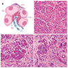
Figure 1
Figure 1. The normal and injured pancreas
(A) Schematic diagram of an acinar unit, indicating acinar cells (AC) with apically-localized zymogen granules (ZG), centroacinar cells (CAC) and duct cells (DC), and quiescent pancreatic stellate cell (psc). (B) Hematoxylin-and-eosin (H&E) stained section of a normal human pancreas, with an exemplary acinar structure outlined with dotted line. (C–D) H&E stained sections of human chronic pancreatitis specimens of varying severity, indicating fibrosis (FI) as well as examples of putative acinar-ductal metaplasia (ADM).
Figure 2
Figure 2. Caerulein-induced pancreatitis and metaplasia
Sections from mice injected with saline solution as a negative control (left), or with high doses of caerulein to induce pancreatitis and harvested 2 days (middle) or 14 days (right) post-injection, stained with H&E (A–C) or by immunohistochemistry against the duct marker cytokeratin-19 (D–F, brown). Metaplasia (open arrowhead) and inflammatory cell infiltration (closed arrowhead) are widespread at 2 days, accompanied by upregulation of CK19 in acinar cells, but are essentially undetectable when regeneration is complete at 14 days. Figure adapted from ref. (46), under a Creative Commons Attribution (CC-BY) 3.0 License (copyright held by L.C.M. and M.K.).
Figure 3
Figure 3. Model for exocrine pancreas injury and regeneration
In response to initial injury, acinar cells recruit leukocytes via NFκB-dependent cytokines, and leukocytes further stimulate NFκB in acinar cells to amplify injury and induce cell death and acinar-ductal metaplasia (ADM) (how individual cells choose between death and ADM is unknown). Dedifferentiated acinar cells continue to recruit and activate inflammatory cells, which in turn continue to stimulate acinar cell NFκB signaling. Pancreatic stellate cells (PSCs) are also activated by leukocytes and may produce additional pro-metaplasia signals, although the role of PSCs in vivo remains uncertain. In acute injury, developmental factors are activated to terminate this loop and induce redifferentiation, while chronic injury results when metaplasia cannot be resolved.

References

    1. Steinberg WM. Acute Pancreatitis. In: Sleisenger MH, Feldman M, Friedman LS, Brandt LJ, editors. Sleisenger & Fordtran’s gastrointestinal and liver disease: pathophysiology, diagnosis, management. Philadelphia: Saunders; 2006.
    1. Pan FC, Wright C. Pancreas organogenesis: From bud to plexus to gland. Dev Dyn. 2011;240:530–65. - PubMed
    1. Shih HP, Wang A, Sander M. Pancreas organogenesis: from lineage determination to morphogenesis. Annu Rev Cell Dev Biol. 2013;29:81–105. - PubMed
    1. Kretzschmar K, Watt FM. Lineage tracing. Cell. 2012;148:33–45. - PubMed
    1. Pan FC, Bankaitis ED, Boyer D, Xu X, Van de Casteele M, Magnuson MA, Heimberg H, Wright CV. Spatiotemporal patterns of multipotentiality in Ptf1a-expressing cells during pancreas organogenesis and injury-induced facultative restoration. Development. 2013;140:751–64. - PMC - PubMed

Publication types

LinkOut - more resources