Review
. 2015:10:173-94.
doi: 10.1146/annurev-pathol-012513-104649. Epub 2014 Oct 27.

The role of endoplasmic reticulum stress in human pathology

Affiliations
Review

The role of endoplasmic reticulum stress in human pathology

Scott A Oakes et al. Annu Rev Pathol. 2015.

Abstract

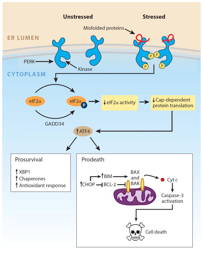
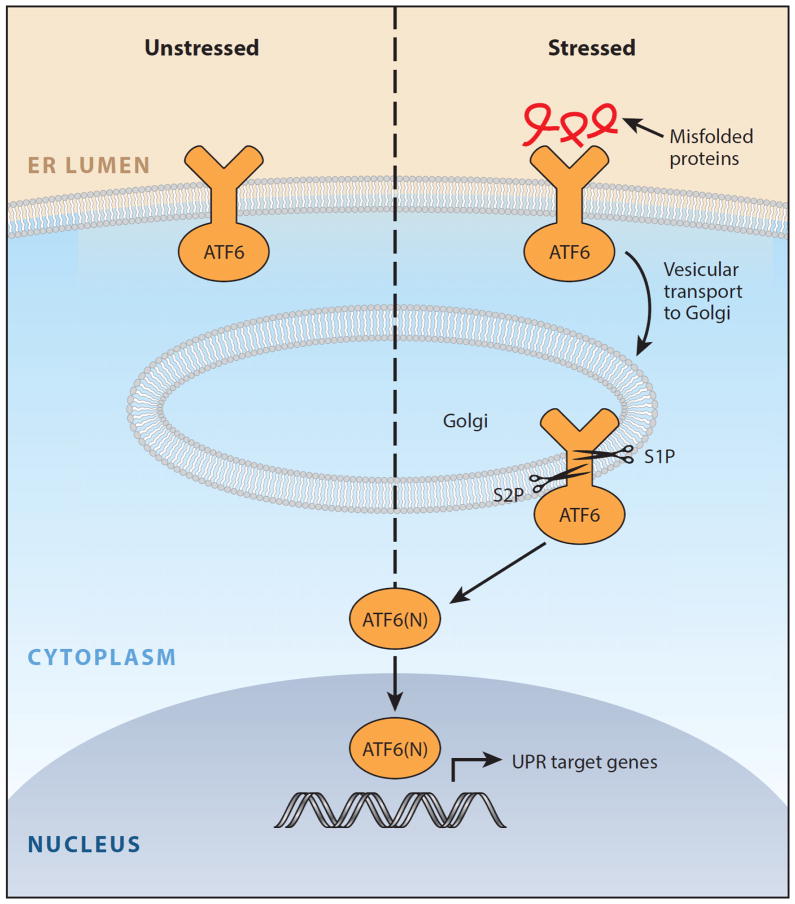
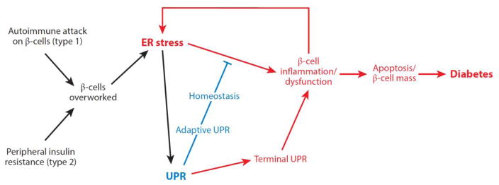
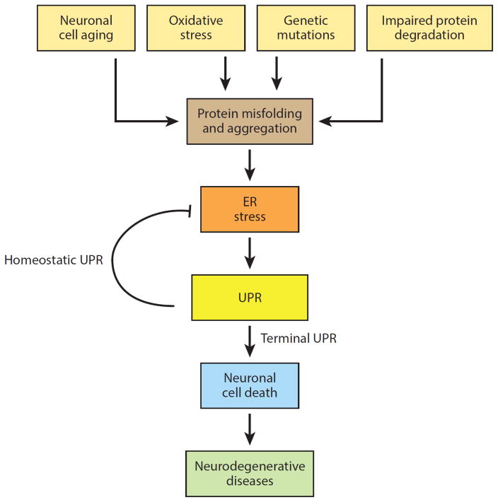
Numerous genetic and environmental insults impede the ability of cells to properly fold and posttranslationally modify secretory and transmembrane proteins in the endoplasmic reticulum (ER), leading to a buildup of misfolded proteins in this organelle--a condition called ER stress. ER-stressed cells must rapidly restore protein-folding capacity to match protein-folding demand if they are to survive. In the presence of high levels of misfolded proteins in the ER, an intracellular signaling pathway called the unfolded protein response (UPR) induces a set of transcriptional and translational events that restore ER homeostasis. However, if ER stress persists chronically at high levels, a terminal UPR program ensures that cells commit to self-destruction. Chronic ER stress and defects in UPR signaling are emerging as key contributors to a growing list of human diseases, including diabetes, neurodegeneration, and cancer. Hence, there is much interest in targeting components of the UPR as a therapeutic strategy to combat these ER stress-associated pathologies.

Keywords: apoptosis; cancer; diabetes; neurodegeneration; protein misfolding; unfolded protein response.


Figures

Figure 1
Figure 1
IRE1α arm of the unfolded protein response. IRE1α is an endoplasmic reticulum (ER) transmembrane protein that becomes activated when misfolded proteins in the ER lumen bind to its luminal domains. The cytoplasmic tail of IRE1α has two enzymatic activities—a serine/threonine kinase domain and an endoribonuclease (RNase) domain. Upon luminal binding of misfolded proteins, IRE1α’s kinase becomes activated and trans-autophosphorylates multiple serine/threonine residues on the cytosolic tail. IRE1α phosphorylation leads to allosteric activation of the adjacent RNase. The consequences of IRE1α activation vary depending on the level of ER stress. In response to low levels of ER stress, IRE1α’s RNase excises a 26-nt intron from the mRNA encoding the XBP1 (X-box protein 1) transcription factor to produce the homeostatic transcription factor XBP1s. XBP1s then translocates to the nucleus and induces transcription of many genes that augment ER size and function in an attempt to restore ER homeostasis. However, if ER stress is irremediable, IRE1α becomes hyperactivated and undergoes homo-oligomerization. Under sustained oligomerization, IRE1α’s RNase endonucleolytically degrades hundreds of ER-localized mRNAs containing an N-terminal signal sequence, which depletes ER cargo and protein-folding components to further worsen ER stress. Moreover, when hyperactivated, IRE1α’s RNase directly cleaves select microRNAs that normally repress proapoptotic targets. In addition to signaling through RNA substrates, IRE1α oligomerization has been shown to induce activation or upregulation of a number of proinflammatory proteins, including the pro-oxidant protein TXNIP (thioredoxin-interacting protein), to activate the inflammasome and its Caspase-1-dependent prodeath pathway. Finally, sustained IRE1α oligomerization serves as an activation platform for ASK1 (apoptosis signal–regulating kinase) and its downstream target JNK (c-Jun NH2-terminal kinase). Phosphorylation by JNK has been reported to both activate proapoptotic BIM and inhibit antiapoptotic BCL-2. Once activated, BH3-only proteins such as BID and BIM disable mitochondrial protecting proteins (e.g., BCL-2, BCL-XL, MCL-1) and in some cases directly trigger the multidomain proapoptotic BAX and BAK proteins to permeabilize the outer mitochondrial membrane and release toxic mitochondrial proteins, such as cytochrome c, into the cytoplasm, where they lead to activation of downstream effector caspases (e.g., Caspase-3) and cell demise. Modified with permission from Reference . Copyright © 2014 by Elsevier.
Figure 2
Figure 2
PERK arm of the unfolded protein response. PERK is an endoplasmic reticulum (ER) transmembrane protein that contains a single cytosolic kinase. When its luminal domains are dimerized in the presence of misfolded proteins, PERK phosphorylates eukaryotic translation initiation factor 2α (eIF2α). Phosphorylation inhibits eIF2α activity and hence slows down global protein translation, giving the cell extra time to attempt to fold the backlog of proteins already present in the ER lumen. In contrast, translation of the transcription factor ATF4 (activating transcription factor 4) is selectively upregulated when the amount of active eIF2α is limiting. ATF4 expression transcriptionally upregulates CHOP (C/EBP-homologous protein; also known as GADD153), which tips the ER toward homeostasis through induction of a number of corrective genes, including XBP1 and chaperones. Although a temporary pause in protein translation due to eIF2α phosphorylation can be beneficial for cells under ER stress, a protracted block in translation from sustained PERK signaling is incompatible with survival. Moreover, high levels of CHOP/GADD153 transcription factor can inhibit the expression of antiapoptotic BCL-2 to hasten cell death and upregulate proapoptotic BIM to trigger activation of the mitochondria-dependent apoptotic pathway. Modified with permission from Reference . Copyright © 2014 by Elsevier.
Figure 3
Figure 3
ATF6 arm of the unfolded protein response. In the presence of misfolded proteins, ATF6 translocates to the Golgi and is cleaved by the Site-1 and Site-2 proteases to release the ATF6(N) transcription factor contained within its cytoplasmic tail. Together with XBP1s, ATF6(N) increases transcription of targets that expand endoplasmic reticulum (ER) size and increase its protein-folding capacity to promote cell survival. Modified with permission from Reference . Copyright © 2014 by Elsevier.
Figure 4
Figure 4
Model for the role of endoplasmic reticulum (ER) stress in diabetes. ER stress is emerging as an important form of β-cell injury in both type 1 and type 2 diabetes. Overwork of β-cells under conditions of insulin resistance, such as secondary to obesity, may promote attrition of β-cells through unfolded protein response (UPR)-mediated apoptosis in type 2 diabetes. Similarly, as the islet becomes inflamed under autoimmune attack in type 1 diabetes, the per-cell workload of the remaining β-cells to secrete proinsulin increases. Taken together, these combined insults may lead to critically enhanced ER stress in the remaining β-cells, thus hastening their demise through a vicious cycle that ultimately leads to diabetes. Modified with permission from Reference . Copyright © 2014 by Elsevier.
Figure 5
Figure 5
Model for the role of endoplasmic reticulum (ER) stress in neurodegeneration. In addition to rare inherited mutations in a single protein that disrupt its proper folding, degenerating neurons are exposed to numerous other insults (e.g., oxidative stress, inflammation, metabolic disturbances) that can compromise protein folding and lead to ER stress. Abbreviation: UPR, unfolded protein response. Modified with permission from Reference . Copyright © 2014 by Elsevier.

References

    1. Fagone P, Jackowski S. Membrane phospholipid synthesis and endoplasmic reticulum function. J Lipid Res. 2009;50(Suppl):S311–16. - PMC - PubMed
    1. Anelli T, Sitia R. Protein quality control in the early secretory pathway. EMBO J. 2008;27:315–27. - PMC - PubMed
    1. Tu BP, Weissman JS. Oxidative protein folding in eukaryotes: mechanisms and consequences. J Cell Biol. 2004;164:341–46. - PMC - PubMed
    1. Sevier CS, Kaiser CA. Formation and transfer of disulphide bonds in living cells. Nat Rev Mol Cell Biol. 2002;3:836–47. - PubMed
    1. van Anken E, Braakman I. Versatility of the endoplasmic reticulum protein folding factory. Crit Rev Biochem Mol Biol. 2005;40:191–228. - PubMed

MeSH terms