Review
. 2014 Nov 7;4(4):621-65.
doi: 10.3390/life4040621.

Microgravity-induced fluid shift and ophthalmic changes

Affiliations
Review

Microgravity-induced fluid shift and ophthalmic changes

Emily S Nelson et al. Life (Basel). .

Abstract

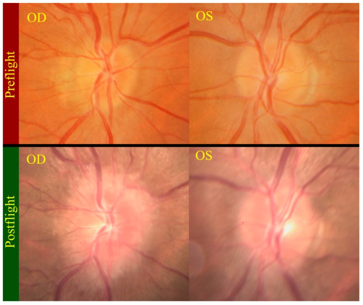
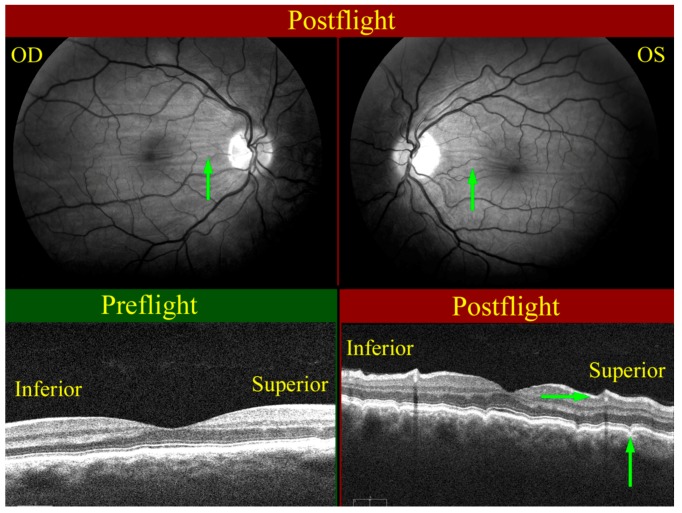
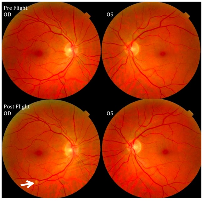
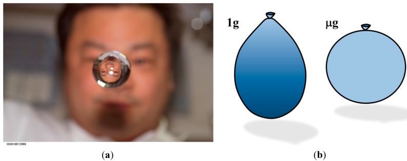
Although changes to visual acuity in spaceflight have been observed in some astronauts since the early days of the space program, the impact to the crew was considered minor. Since that time, missions to the International Space Station have extended the typical duration of time spent in microgravity from a few days or weeks to many months. This has been accompanied by the emergence of a variety of ophthalmic pathologies in a significant proportion of long-duration crewmembers, including globe flattening, choroidal folding, optic disc edema, and optic nerve kinking, among others. The clinical findings of affected astronauts are reminiscent of terrestrial pathologies such as idiopathic intracranial hypertension that are characterized by high intracranial pressure. As a result, NASA has placed an emphasis on determining the relevant factors and their interactions that are responsible for detrimental ophthalmic response to space. This article will describe the Visual Impairment and Intracranial Pressure syndrome, link it to key factors in physiological adaptation to the microgravity environment, particularly a cephalad shifting of bodily fluids, and discuss the implications for ocular biomechanics and physiological function in long-duration spaceflight.


Figures

Figure 1
Figure 1
Reference location of Optic Nerve Sheath Diameter (ONSD) measurement for determining risk of elevated Intracranial Pressure (ICP).
Figure 2
Figure 2
Remotely guided on-orbit ultrasound images showing the OD (right eye) Optic Nerve Sheath Diameter (ONSD, green outline) distended to approximately 12 mm, thus suggesting increased Intracranial Pressure (ICP). Comparing the optic nerve (pink outline) of the right eye with the left (OS) eye, a kink can clearly be seen in the OD optic nerve. Source: NASA Longitudinal Study of Astronaut Health.
Figure 3
Figure 3
The postflight magnetic resonance image on the left shows that optic nerve kinking is present, while the control orbit on the right does not exhibit any tortuosity in the nerve. Source: National Aeronautics and Space Administration.
Figure 4
Figure 4
(a) Inflight ultrasound of a globe showing flattening of the posterior globe; (b) Magnetic resonance image (MRI) of a normal globe before long-duration spaceflight; and (c) MRI of a globe after a long-duration flight exhibiting posterior flattening. Source: NASA Longitudinal Study of Astronaut Health.
Figure 5
Figure 5
Postflight fundus examination photos (bottom) show grade 3 and grade 1 optic disc edema in the right (OD) and left (OS) optic discs respectively. The top photos show the preflight fundus examination of normal optic discs. Source: NASA Longitudinal Study of Astronaut Health.
Figure 6
Figure 6
Choroidal folds (green arrows) as seen on the right (OD, upper left), and left (OS, upper right) globes. The wavy pattern (highlighted by green arrows) in the choroidal/retinal layer shown in the OCT image taken post-flight (bottom right) relative to the pre-flight OCT image (bottom left) exhibits the presence of choroidal folds. Source: NASA Longitudinal Study of Astronaut Health.
Figure 7
Figure 7
Pre- and postflight fundus examination images of the first Visual Impairment and Intracranial Pressure (VIIP) case. As identified by the white arrow in the right eye, a single cotton-wool spot was discovered. Source: National Aeronautics and Space Administration.
Figure 8
Figure 8
(a) Astronaut Leroy Chiao observes his reflected and refracted image in a spherical droplet of water onboard the ISS. Source: National Aeronautics and Astronautics Association [66]; (b) The equilibrium shapes for a water-filled balloon in 1 g and microgravity (μg).
Figure 9
Figure 9
(a) Schematic of a hypothetical human-shaped balloon filled with water in an erect posture; and (b) in Head-Down Tilt; (c) tube-shaped balloon in HDT; (d) human-shaped balloon in microgravity.
Figure 10
Figure 10
(a) Pressure/volume relation for the human eye, adapted from [75]; and (b) Volume/pressure relation for the entire cerebrospinal fluid space, adapted from [74].
Figure 11
Figure 11
Effect of microgravity on fluid distribution in the human body during the 84-day Skylab 4 mission. (a) Measurement locations. The second column shows the change in height and circumference of the waist and chest on inspiration (insp) and expiration (exp) for the (b) commander; (c) science pilot; and (d) pilot. The third column shows left limb net volume change for the (e) commander; (f) science pilot; and (g) pilot. After Thornton [43].
Figure 12
Figure 12
Number of major blood vessels in choroidal (a) inflow (data obtained from [202]) and (b) outflow (data obtained from [204]).

References

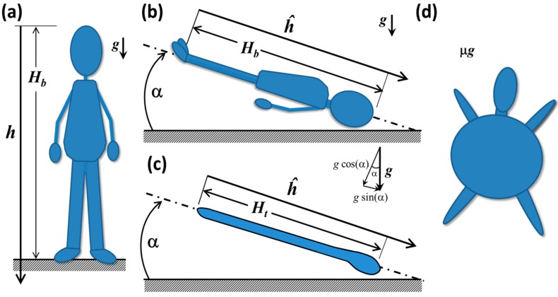
    1. This cephalad shifting of fluid from the extremities toward the head is often referred to as the “cephalad fluid shift” in the spaceflight community.

    1. LeBlanc A., Schneider V., Shackelford L., West S., Oganov V., Bakulin A., Voronin L. Bone mineral and lean tissue loss after long duration space flight. J. Musculoskelet. Neuronal Interact. 2000;1:157–160. - PubMed
    1. LeBlanc A., Lin C., Shackelford L., Sinitsyn V., Evans H., Belichenko O., Schenkman B., Kozlovskaya I., Oganov V., Bakulin A., et al. Muscle volume, MRI relaxation times (T2), and body composition after spaceflight. J. Appl. Physiol. 2000;89:2158–2164. - PubMed
    1. Blaber A.P., Goswami N., Bondar R.L., Kassam M.S. Impairment of cerebral blood flow regulation in astronauts with orthostatic intolerance after flight. Stroke. 2011;42:1844–1850. doi: 10.1161/STROKEAHA.110.610576. - DOI - PubMed
    1. Clement G., Ngo-Anh J.T. Space physiology II: Adaptation of the central nervous system to space flight--past, current, and future studies. Eur. J. Appl. Physiol. 2013;113:1655–1672. doi: 10.1007/s00421-012-2509-3. - DOI - PubMed