

The value of trichoscopy in the differential diagnosis of scalp lesions in pemphigus vulgaris and pemphigus foliaceus
- PMID: 25387515
- PMCID: PMC4230679
- DOI: 10.1590/abd1806-4841.20143830
The value of trichoscopy in the differential diagnosis of scalp lesions in pemphigus vulgaris and pemphigus foliaceus
Abstract
Background: Trichoscopy is becoming increasingly popular in diagnosing hair and scalp diseases. Scalp involvement in pemphigus is common. The scalp may be the first or only site of clinical manifestation of the disease.
Objective: The aim of this study was to analyze whether trichoscopy may be useful in aiding differential diagnosis of scalp lesions in patients with pemphigus vulgaris and pemphigus foliaceus.
Methods: Trichoscopy was performed in 19 patients with scalp lesions in the course of pemphigus (9 patients with pemphigus vulgaris and 10 with pemphigus foliaceus). In all patients, the diagnosis of scalp pemphigus was confirmed by histopathology. The working magnification was 20-fold and 70-fold.
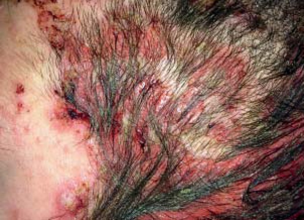
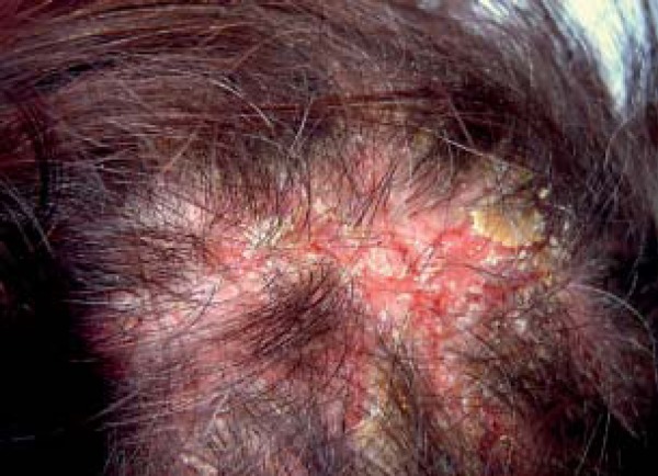
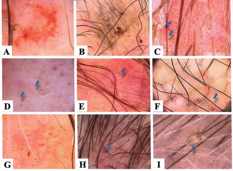
Results: The most frequently observed trichoscopy features of pemphigus lesions were: extravasations (18/19; 94.7%) and yellow hemorrhagic crusts (11/19; 57.9%). Yellow dots with whitish halo were observed in 6/19 (31.6%) patients with pemphigus. White polygonal structures were observed in pemphigus foliaceus (6/10; 60%), but not in pemphigus vulgaris. Vascular abnormalities were more frequent in pemphigus vulgaris, when compared to pemphigus foliaceus, and were associated with a severe course of disease. Linear serpentine vessels were the most frequent vascular abnormality in patients with pemphigus vulgaris and pemphigus foliaceus (77.8% and 30%, respectively).
Conclusion: Trichoscopy may serve as a useful supplementary method in the differential diagnosis of pemphigus, especially in cases of desquamative or exudative lesions limited to the scalp. Extravasations, yellow hemorrhagic crusts, yellow dots with whitish halo, white polygonal structures and linear serpentine vessels are trichoscopy features which may suggest the diagnosis of pemphigus.
Conflict of interest statement
Conflict of interest: None
Figures
Similar articles
-
The Significance of Scalp Involvement in Pemphigus: A Literature Review.Biomed Res Int. 2018 Mar 25;2018:6154397. doi: 10.1155/2018/6154397. eCollection 2018. Biomed Res Int. 2018. PMID: 29770335 Free PMC article. Review.
-
Trichoscopy - a useful tool in the preliminary differential diagnosis of autoimmune bullous diseases.Int J Dermatol. 2017 Oct;56(10):996-1002. doi: 10.1111/ijd.13725. Epub 2017 Aug 30. Int J Dermatol. 2017. PMID: 28856676
-
Diagnostic Accuracy of Trichoscopy in Inflammatory Scalp Diseases: A Systematic Review.Dermatology. 2022;238(3):412-421. doi: 10.1159/000517516. Epub 2021 Jul 15. Dermatology. 2022. PMID: 34265772
-
Pemphigus scalp lesions: is trichoscopy helpful in clinical diagnosis?Int J Dermatol. 2021 Jan;60(1):81-87. doi: 10.1111/ijd.15231. Epub 2020 Oct 5. Int J Dermatol. 2021. PMID: 33017059
-
Miniaturization of sebaceous glands: A novel histopathological finding in pemphigus vulgaris and pemphigus foliaceus of the scalp.J Cutan Pathol. 2017 Oct;44(10):835-842. doi: 10.1111/cup.12994. Epub 2017 Jul 21. J Cutan Pathol. 2017. PMID: 28632917
Cited by
-
Tubular Hair Casts in Trichoscopy of Hair and Scalp Disorders.Int J Trichology. 2019 Jan-Feb;11(1):14-19. doi: 10.4103/ijt.ijt_77_18. Int J Trichology. 2019. PMID: 30820128 Free PMC article.
-
Trichoscopy of Pemphigus Vulgaris and Foliaceus on Black Scalp.Skin Appendage Disord. 2025 Apr;11(2):147-150. doi: 10.1159/000540968. Epub 2024 Oct 15. Skin Appendage Disord. 2025. PMID: 40176993
-
Scalp Erosions that Do Not Heal: The Diagnostic Value of Trichoscopy.Skin Appendage Disord. 2021 Feb;7(2):155-158. doi: 10.1159/000513135. Epub 2021 Jan 27. Skin Appendage Disord. 2021. PMID: 33796565 Free PMC article. No abstract available.
-
The Significance of Scalp Involvement in Pemphigus: A Literature Review.Biomed Res Int. 2018 Mar 25;2018:6154397. doi: 10.1155/2018/6154397. eCollection 2018. Biomed Res Int. 2018. PMID: 29770335 Free PMC article. Review.
-
Avian-Inspired Analogies in Dermatology.Indian Dermatol Online J. 2025 Apr 15;16(3):435-439. doi: 10.4103/idoj.idoj_1039_24. eCollection 2025 May-Jun. Indian Dermatol Online J. 2025. PMID: 40395580 Free PMC article. No abstract available.
References
-
- Cunha PR, Barraviera SR. Autoimmune bullous dermatoses. An Bras Dermatol. 2009;84:111–124. - PubMed
-
- Ruocco V, Ruocco E, Lo Schiavo A, Brunetti G, Guerrera LP, Wolf R. Pemphigus: etiology, pathogenesis, and inducing or triggering factors: facts and controversies. Clin Dermatol. 2013;31:374–381. - PubMed
-
- Sar-Pomian M, Kolacinska-Strasz Z, Labecka H, Krainska-Wojcik T, Olszewska M. Scalp lesions in pemphigus. Przegl Dermatol. 2010;97:14–20.
-
- Oretti G, Giordano D, Di Lella F, Gradoni P, Zendri E, Ferri T. Unilesional pemphigus vulgaris of the scalp after cochlear implantation. Am J Otolaryngol. 2011;32:80–81. - PubMed
Publication types
MeSH terms
Substances
LinkOut - more resources
Full Text Sources
Other Literature Sources
Medical