Review
. 2014 Dec;47(12):655-9.
doi: 10.5483/bmbrep.2014.47.12.241.

Integrin activation

Affiliations
Review

Integrin activation

Mark H Ginsberg. BMB Rep. 2014 Dec.

Abstract

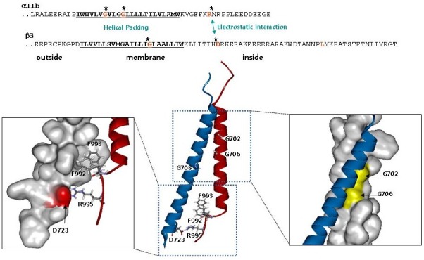
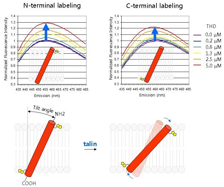
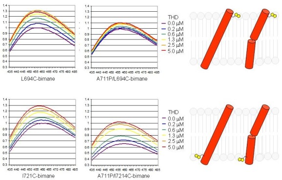
Integrin-mediated cell adhesion is important for development, immune responses, hemostasis and wound healing. Integrins also function as signal transducing receptors that can control intracellular pathways that regulate cell survival, proliferation, and cell fate. Conversely, cells can modulate the affinity of integrins for their ligands a process operationally defined as integrin activation. Analysis of activation of integrins has now provided a detailed molecular understanding of this unique form of "inside-out" signal transduction and revealed new paradigms of how transmembrane domains (TMD) can transmit long range allosteric changes in transmembrane proteins. Here, we will review how talin and mediates integrin activation and how the integrin TMD can transmit these inside out signals.


Figures

Fig. 1.
Fig. 1.. Structure of integrin αIIbβ3 TMD (ribbon view; αIIb in red and β3 in blue. From PDB 2K9J) showing the two interaction interfaces. Right, outer membrane clasp (OMC) illustrating the helical packing involving αIIb Gly 702 and 706. Left, inner membrane clasp (IMC) showing the electrostatic interaction between αIIb Arg995 and β3 Asp723. Also depicted are the hydrophobic interactions of αIIb Phe992,933 with the β3 TMD. Adapted from reference .
Fig. 2.
Fig. 2.. Talin changes the topology of the β3 TMD. A peptide containing the β3 TMD and cytoplasmic domain was labeled with environment sensitive bimanes at the outer edge of the TMD (N terminal labeling, Leu694) or at the TMD cytosol interface (C-terminal labeling, Ile721). The peptides were individually embedded in phospholipid nanodiscs and increasing concentrations of talin head domain (THD) were added and bimanes emission spectra were recorded. The increased fluorescence indicates that both sides of the β3 TMD were in a less polar environment suggesting that THD increased the tilting of the β3 TMD. Adapted from reference .
Fig. 3.
Fig. 3.. A proline kink prevents transmission of altered tilt across the β3 TMD. In the left two panels The experimental design was identical to that in Fig. 2 and depicts the talin-induced increased embedding at both the inner (where THD binds) and outer edges of the TMD. Introduction of a flexible kink by β3 (A711P) mutation (right two panels) prevents the transmission of increased embedding of the inner TMD to the outer region. Adapted from reference .

References

    1. Hynes R. O. Integrins: bidirectional, allosteric signaling machines. Cell. (2002);110:673–687. doi: 10.1016/S0092-8674(02)00971-6. - DOI - PubMed
    1. Giancotti F. G., Ruoslahti E. Integrin signaling. Science. (1999);285:1028–1032. doi: 10.1126/science.285.5430.1028. - DOI - PubMed
    1. Kim C., Ye F., Ginsberg M. H. Regulation of integrin activation. Annu. Rev. Cell. Dev. Biol. (2011);27:321–345. doi: 10.1146/annurev-cellbio-100109-104104. - DOI - PubMed
    1. Shattil S. J., Kim C., Ginsberg M. H. The final steps of integrin activation: the end game. Nat. Rev. Mol. Cell. Biol. (2010);11:288–300. doi: 10.1038/nrm2871. - DOI - PMC - PubMed
    1. Shattil S. J., Newman P. J. Integrins: dynamic scaffolds for adhesion and signaling in platelets. Blood. (2004);104:1606–1615. doi: 10.1182/blood-2004-04-1257. - DOI - PubMed