

doi: 10.1007/978-1-4939-1568-2_26.
Evaluation of synovial mast cell functions in autoimmune arthritis
Affiliations
- PMID: 25388266
- PMCID: PMC6817810
- DOI: 10.1007/978-1-4939-1568-2_26
Item in Clipboard
Evaluation of synovial mast cell functions in autoimmune arthritis
Methods Mol Biol.
2015.
Abstract
Mast cells are innate immune effector cells that reside in the healthy synovial sublining and expand in number with inflammation. These cells can play an important role in initiation of arthritis, but much about their biology and importance remains obscure. This chapter reviews the use of animal models for the study of mast cells in arthritis, with a particular focus on the K/BxN serum transfer model. We discuss tissue preparation and histological analysis for the assessment of joint inflammation, injury, and the presence and phenotype of synovial mast cells, as well as the use of bone marrow-derived mast cell (BMMC) engraftment into W/Wv mice as a tool to isolate the role of mast cells in joint inflammation and injury.
Figures
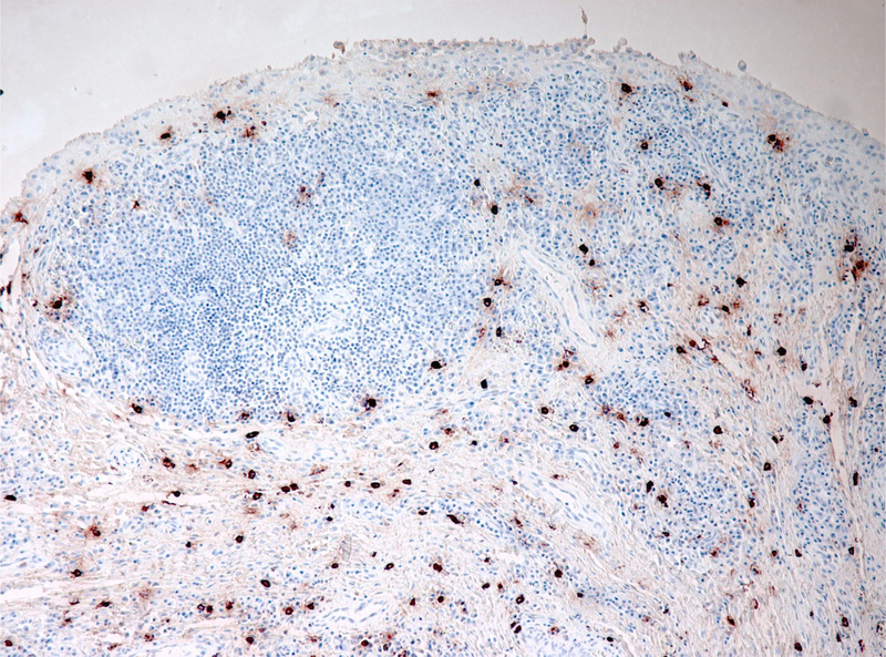
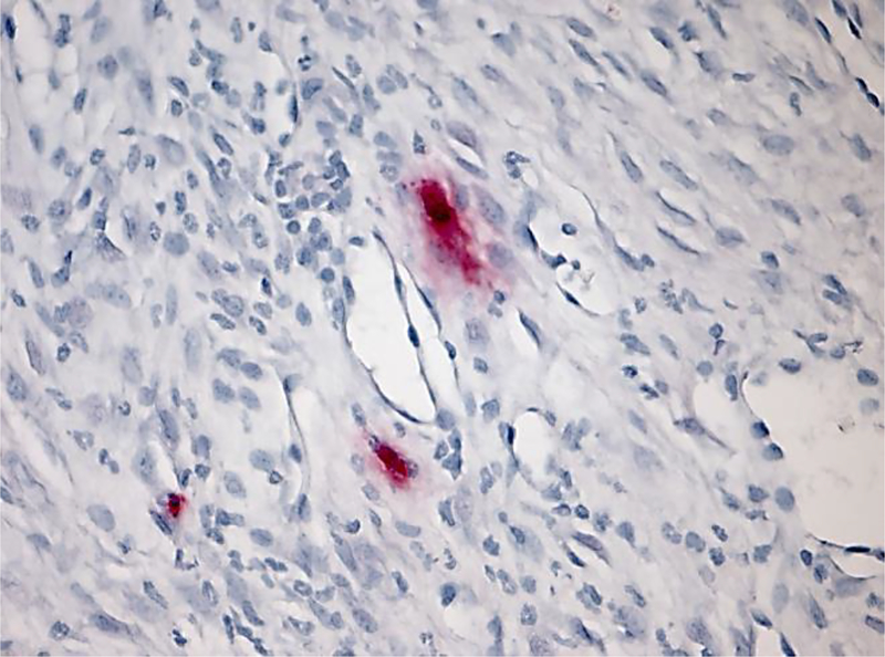
Human RA synovium stained for tryptase (red) highlights abundance of mast cells in chronically inflamed joint tissue; reproduced from reference (2).
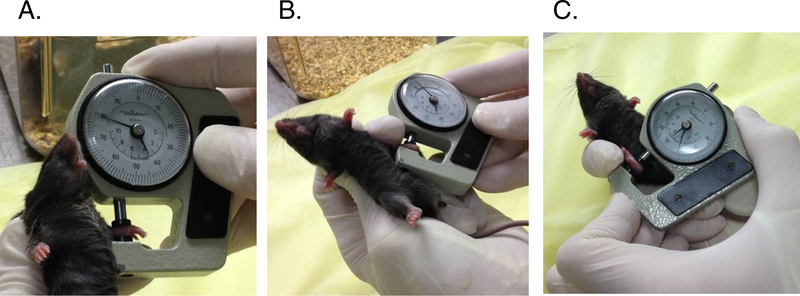
Measurement of paw thickness in arthritis. A. Forepaw measurement in dorsal-ventral axis. B. Left hindpaw measurement. C. Right hindpaw measurement. Note in B and C that a finger in the restraining hand is used to maintain the measured joint in a degree of flexion that must be maintained constant from measurement to measurement. The caliper must be held in each hand alternately.
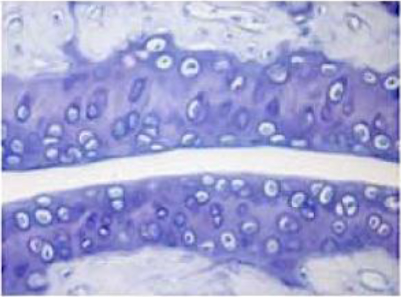
Cartilage proteoglycan staining with toluidine blue. This murine tibotalar joint was sectioned and stained with toluidine blue. Proteoglycan within cartilage is stained as darker bluish-purple.
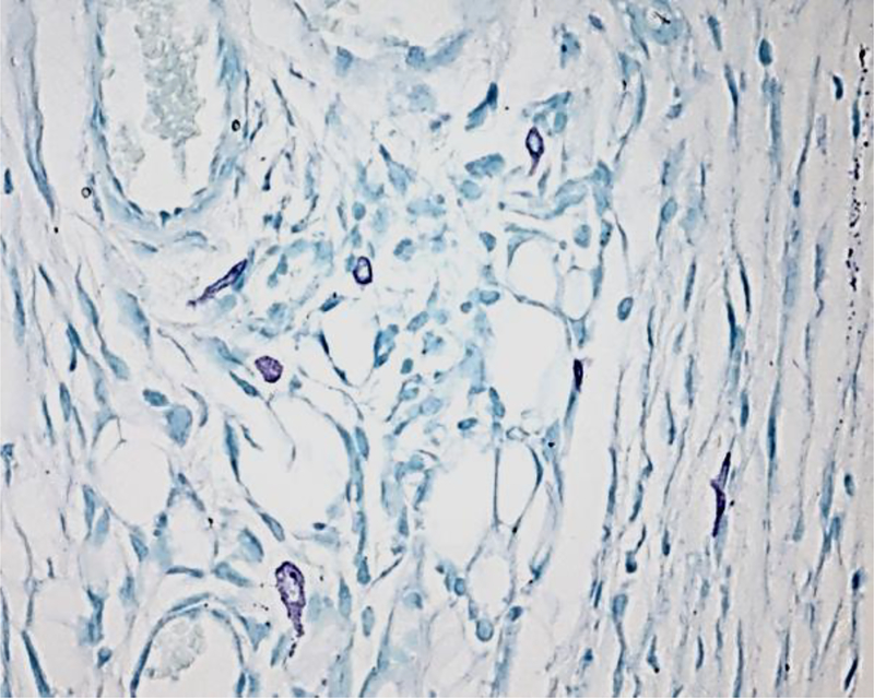
Toluidine blue staining of synovial mast cells. Note purple color of mast cells compared to blue background.
CAE staining of synovial mast cells
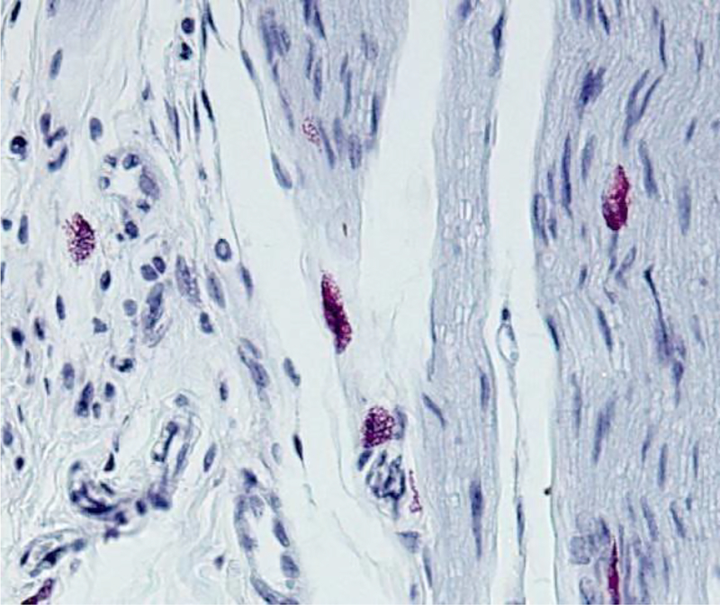
Immunohistochemistry of murine synovial mast cells for mMCP6.
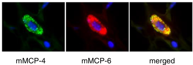
Immunofluorescence staining of murine synovial mast cells for mMCP4 (green) and −6 (red). Merged figure (yellow) shows that both proteases are expressed in the synovial mast cell.
References
-
- Castor W. The microscopic structure of normal human synovial tissue. Arthritis Rheum 1960;3:140–151. - PubMed
-
- Nigrovic PA, Lee DM. Synovial mast cells: role in acute and chronic arthritis. Immunol Rev 2007;217:19–37. - PubMed
-
- Crisp AJ, Chapman CM, Kirkham SE, Schiller AL, Krane SM. Articular mastocytosis in rheumatoid arthritis. Arthritis Rheum 1984;27(8):845–51. - PubMed
-
- Monach PA, Benoist C, Mathis D. The role of antibodies in mouse models of rheumatoid arthritis, and relevance to human disease. Adv Immunol 2004;82:217–48. - PubMed
-
- Kouskoff V, Korganow AS, Duchatelle V, Degott C, Benoist C, Mathis D. Organ-specific disease provoked by systemic autoimmunity. Cell 1996;87(5):811–22. - PubMed
Publication types
MeSH terms
Substances
Grants and funding
LinkOut - more resources
Full Text Sources
Other Literature Sources
Medical
