. 2015:1235:179-202.
doi: 10.1007/978-1-4939-1785-3_15.

Advanced imaging and tissue engineering of the human limbal epithelial stem cell niche

Affiliations

Advanced imaging and tissue engineering of the human limbal epithelial stem cell niche

Isobel Massie et al. Methods Mol Biol. 2015.

Abstract

The limbal epithelial stem cell niche provides a unique, physically protective environment in which limbal epithelial stem cells reside in close proximity with accessory cell types and their secreted factors. The use of advanced imaging techniques is described to visualize the niche in three dimensions in native human corneal tissue. In addition, a protocol is provided for the isolation and culture of three different cell types, including human limbal epithelial stem cells from the limbal niche of human donor tissue. Finally, the process of incorporating these cells within plastic compressed collagen constructs to form a tissue-engineered corneal limbus is described and how immunohistochemical techniques may be applied to characterize cell phenotype therein.


Figures
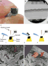
Fig. 1
Fig. 1
Serial block face scanning electron microscopy. (a) Resin block attached to the cryopin, ready to be loaded into the SEM. The dashed white line outlines the surface of the resin block measuring approximately 0.5 × 0.5 mm. (b) Low magnification scanning electron micrograph of the resin block in a. (c) Schematic showing the principle of the SBFSEM (3View). 1: Imaging of the surface of the resin block. 2: A diamond knife inside microscope chamber cuts an ultrathin section away from the specimen. 3: The freshly exposed edge is imaged. (d) Manual segmentation of the area of interest. The nuclei of a stromal cell and epithelial cell are outlined. (e) 3D reconstruction of the areas manually segmented in d. Scale bars: b: 100 μm and d: 5 μm
Fig. 2
Fig. 2
hLEC cultures. (a) hLEC form colonies (left) between the feeder cells (right). (b) hLEC display typical cobblestone morphology, with scant cytoplasm. Scale bars: 200 μm
Fig. 3
Fig. 3
hLF cultures. (a) hLF in culture at sub-confluency. (b) A confluent hLF layer, ready for trypsinization. hLF appear dendritic. Scale bars: 200 μm
Fig. 4
Fig. 4
hCSSC isolation and culture using enzymatic dissociation. (a) Black dotted lines illustrate the limbal region before dissection. (b) Upper half of superficial limbal rim is dissected. (c) hCSSC in culture (passage 1). CSSCs appear small and square in sparsely arranged colonies. Some epithelial colonies (E) and keratocyte- like cells (K) remain visible in early passages. Scale bar: 100 μm
Fig. 5
Fig. 5
RAFT production process. (a) Base of absorbers showing ridged and plain topographies. (b) Guide plate populated with absorbers on top of 24-well plate. (c) Acellular RAFT construct in the base of a 24-well plate. (d) Schematic summarizing the RAFT production process
Fig. 6
Fig. 6
Example of a wholemount-stained RAFT construct. (a) hLEC on RAFT stained for β1-integrin, putative stem cell marker. (b) hLEC on RAFT stained for phalloidin-FITC. (c) Merge. Scale bars: 50 μm
Fig. 7
Fig. 7
Example of a stained RAFT section. (a) hLEC on RAFT stained for p63α, putative stem cell marker. (b) PI counter-staining of hLEC (left) nuclei on RAFT and entrapped hLF (right) nuclei. (c) Merge showing that only basal hLEC express p63α. Scale bars: 20 μm

References

    1. Thoft R, Friend J. The X, Y, Z hypothesis of corneal epithelial maintenance. IOVS. 1983;10:1442–1443. - PubMed
    1. Pellegrini G, Golisano O, Paterna P, et al. Location and clonal analysis of stem cells and their differentiated progeny in the human ocular surface. J Cell Biol. 1999;145:769–782. - PMC - PubMed
    1. Schlotzer-Schrehardt U, Kruse FE. Identification and characterization of limbal stem cells. Exp Eye Res. 2005;81:247–264. - PubMed
    1. Shortt AJ, Secker GA, Munro PM, et al. Characterization of the limbal epithelial stem cell niche: novel imaging techniques permit in vivo observation and targeted biopsy of limbal epithelial stem cells. Stem Cells. 2007;25:1402–1409. - PubMed
    1. Denk W, Horstmann H. Serial block-face scanning electron microscopy to reconstruct three-dimensional tissue nanostructure. PLoS Biol. 2004;2:329. - PMC - PubMed

MeSH terms

LinkOut - more resources