. 2014 Nov 12:12:72.
doi: 10.1186/s12964-014-0072-8.

The E3 ubiquitin ligase RNF121 is a positive regulator of NF-κB activation

Affiliations

The E3 ubiquitin ligase RNF121 is a positive regulator of NF-κB activation

Naima Zemirli et al. Cell Commun Signal. .

Abstract

Background: The nuclear factor κB (NF-κB) family members regulate several biological processes as cell proliferation and differentiation, inflammation, immunity and tumor progression. Ubiquitination plays a key role in NF-κB activation and the ubiquitylated transmitters of the NF-κB signaling cascade accumulate in close proximity to endomembranes.

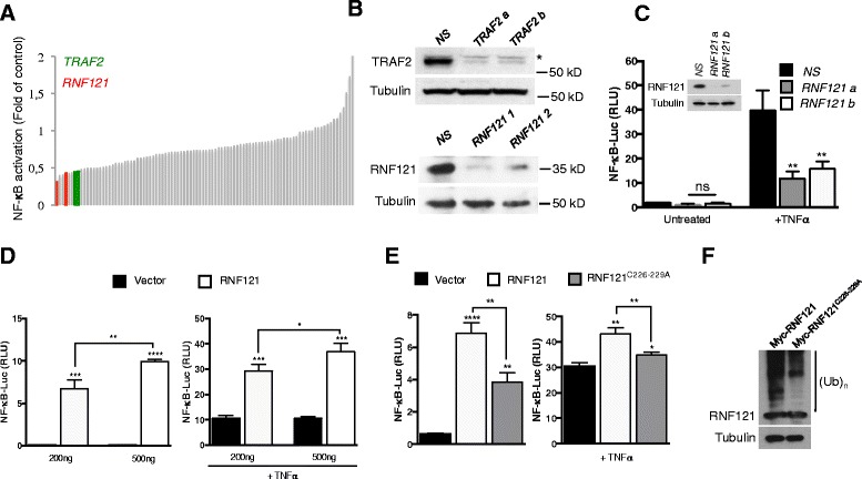
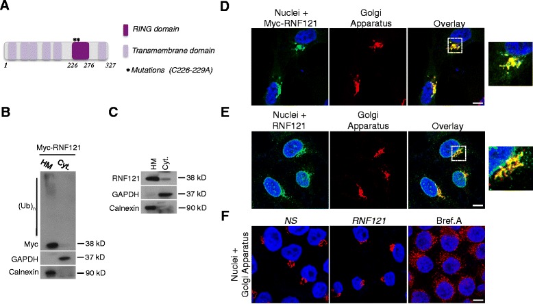
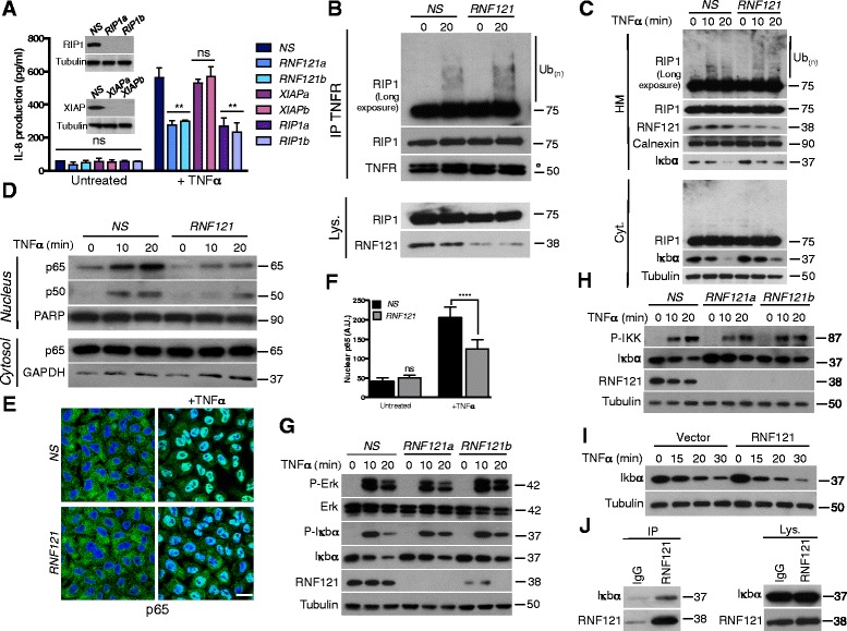
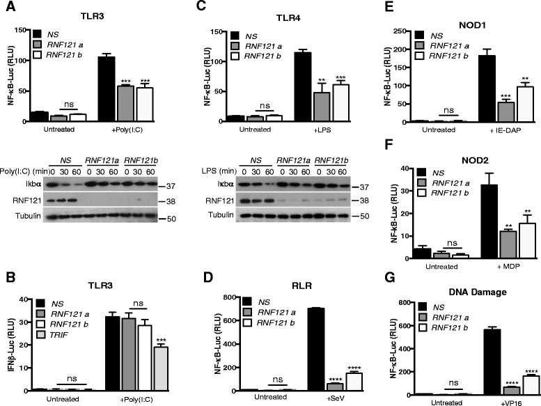
Findings: We performed an unbiased siRNA library screen targeting the 46 E3 ubiquitin ligases bearing transmembrane domains to uncover new modulators of NF-κB activation, using tumor necrosis factor-α (TNF-α) receptor (TNFR) stimulation as a model. We report here the identification of a new Golgi Apparatus-resident protein, RNF121, as an enhancer of NF-κB promoter activity through the catalytic function of its RING domain. From a molecular standpoint, while knocking down RNF121 did not alter RIP1 ubiquitination and IKK activation, the proteasomal degradation of IκBα was impaired suggesting that this E3 ubiquitin ligase regulates this process. However, RNF121 did not directly ubiquitinate IκBα While they were found in the same complex. Finally, we discovered that RNF121 acts as a broad regulator of NF-κB signaling since its silencing also dampens NF-κB activation following stimulation of Toll-Like Receptors (TLRs), Nod-Like Receptors (NLRs), RIG-I-Like Receptors (RLRs) or after DNA damages.

Conclusions: These results unveil an unexpected role of Golgi Apparatus and reveal RNF121 as a new player involved in the signaling leading to NF-κB activation.


Figures

Figure 1
Figure 1
Participation of RNF121 in TNFR-mediated NF-κB activation. (A) NF-κB reporter luciferase assay screen of a siRNA library targeting 46 transmembrane E3 ubiquitin ligases (2 siRNAs/target) in HEK293T cells. Cells were stimulated with TNFα (10 ng/ml) for 6 hrs and fold activation compared to non-specific (NS) siRNA-treated cells was calculated. Red and green histograms indicate siRNA against RNF121 and TRAF2, respectively. TRAF2 was used as a positive control. (B) Cell extracts from HEK293T cells transfected as in (A) were analyzed by immunoblot as indicated. (C) HEK293T cells transfected with a control non-specific (NS) siRNA or with siRNAs against RNF121 (RNF121 a or b), were also transfected 48 hrs later with an NF-κB reporter. 24 hrs later, the cells were either left unstimulated or were stimulated with TNFα (10 ng/ml) for 6 hrs and then were analyzed by luciferase assay. The results were normalized against Renilla luciferase activity [analysis of variance (ANOVA)]. ns: not significant. RLU, Relative Light Units. Inset: Immunoblotting analysis of the knockdown of RNF121 by the specific siRNAs. (D) NF-κB reporter luciferase assay in HEK293T cells transfected with increasing concentrations (200 or 500 ng) of a Myc-tagged plasmid coding for RNF121 and left unstimulated (left panel) or stimulated with TNFα (1 ng/ml) for 6 hrs (right panel) [analysis of variance (Student’s t-tests)]. (E) NF-κB reporter luciferase assay in HEK293T cells transfected with 200 ng of a Myc-tagged plasmid coding for RNF121 or for the mutant RNF121C226-229A and left unstimulated (left panel) or stimulated with TNFα (10 ng/ml) for 6 hrs (right panel) [analysis of variance (Student’s t-tests)]. (F) HEK293T cells were transfected with a Myc-tagged plasmid coding for RNF121 or for the mutant RNF121C226-229A. 24 hrs later, cell extracts were analyzed by immunoblotting. (Ub)n indicates poly-ubiquitylated species.
Figure 2
Figure 2
RNF121 is a Golgi Apparatus-anchored E3 ubiquitin ligase. (A) Scheme showing the main domains of RNF121. The mutations used in Figure 1E and 1F are also indicated. (B) HEK293T cells were transfected with a Myc-tagged plasmid coding for RNF121. 24 hrs later, crude heavy membranes (HM) and cytosolic (Cyt.) fractions were analyzed by immunoblotting as indicated. Calnexin and GAPDH served as purity controls for the HM or cytosolic fraction respectively. (Ub)n indicates poly-ubiquitylated species. (C) Crude heavy membranes (HM) and cytosolic (Cyt.) fractions from HEK293T were analyzed by immunoblotting as indicated. (D) HeLa cells were transfected with a Myc-tagged plasmid coding for RNF121. 24 hrs later, the localization of myc-RNF121 was investigated by immunofluorescence. GM130 was used as a marker for the Golgi Apparatus. Nuclei were stained with DAPI. Representative images are shown, with the boxed areas enlarged on the right. (E) Analysis of the localization of endogenous RNF121 in HeLa cells by immunofluorescence. (F) HeLa were transfected with a control nonspecific siRNA or with a siRNA raised against RNF121. 72 hrs later the Golgi Apparatus morphology was examined. As a control, HeLa cells were treated with Brefeldin A (Bref. A) (50 ng/ml) for 3 hrs. Scale bar : 20 μm.
Figure 3
Figure 3
RNF121 regulates NF-κB activation through IκBα degradation. (A) HEK293T cells were transfected with a control non-specific (NS) siRNA or with the indicated siRNAs. 72 hrs later, the cells were either left untreated or exposed to TNFα (1 ng/ml). The secretion of IL-8 was measured by ELISA. (B) HEK293T cells were transfected for 72 hrs with NS siRNA or with a siRNA against RNF121 (RNF121). Cells were then either left untreated or exposed to 10 ng/ml TNFα for 20 min. Cell lysates (Lys.) were subjected to immunoprecipitation (IP) with an antibody raised against TNFR. ° IgG heavy chains. (C) NS- and RNF121-silenced HEK293T cells were stimulated with TNFα (10 ng/ml) for the indicated times. Crude HM and cytosolic (Cyt.) fractions were analyzed by immunoblotting. (D) Nuclear and cytoplasmic extracts from cells stimulated as in (C) were analyzed by immunoblotting as indicated. (E) NS- and RNF121-silenced HeLa cells were either left untreated or exposed to TNFα for 20 min. Nuclear translocation of the p65 NF-κB subunit was assessed by immunofluorescence. Scale bar : 50 μm. The pixel intensity of the nuclear signal of p65 in each condition was quantified in (F). A.U.: Arbitrary unit. (G) NS- and RNF121-silenced HEK293T were stimulated with TNFα for the indicated times, then were subjected to immunoblotting analysis as indicated. (H) Same conditions as (G) but cells were pre-treated 5 min with the broad phosphatase inhibitor Calyculin A (50 nM) before TNFα stimulation. (I) HEK293T cells were transfected with a control empty vector or Myc-tagged plasmid coding for RNF121. 24 hrs later, cells were stimulated with TNFα for the indicated times and cell extracts were analyzed by immunoblotting as indicated. (J) Cell lysates (Lys.) from HEK293T were subjected to immunoprecipitation (IP) with an antibody raised against RNF121 or with a control IgG.
Figure 4
Figure 4
RNF121 is a broad regulator of NF-κB activation. (A) HEK293T cells stably expressing TLR3 were transfected with a control non-specific (NS) siRNA or with siRNAs against RNF121 (RNF121 a or b). 48 hrs later, the cells were also transfected with an NF-κB reporter. 24 hrs later, the cells were either left unstimulated or were stimulated with poly(I:C) (10 μg/ml) for 6 hrs and then were analyzed by luciferase assay with normalization against Renilla luciferase activity. The NS- and RNF121-silenced cells were also stimulated with poly(I:C) for the indicated times, then were subjected to immunoblotting analysis as indicated. (B) NS-,RNF121- and TRIF-silenced HEK293T stably expressing TL3 were transfected with an IFNβ reporter. 24 hrs later, the cells were either left unstimulated or were stimulated with poly(I:C) (10 μg/ml) for 6 hrs and then were analyzed by luciferase assay. (C) HEK293T cells stably expressing TLR4 were transfected as in (A). Then, cells were either left unstimulated or were stimulated with LPS (10 μg/ml) for 6 hrs and then were analyzed by luciferase assay. The NS- and RNF121-silenced cells were also stimulated with LPS (10 μg/ml) for the indicated times, then were subjected to immunoblotting analysis as indicated. (D) HEK293T cells were transfected as in (A). Then, cells were either left unstimulated or were infected with Sendai virus (SeV) for 6 hrs and then were analyzed by luciferase assay. HEK293T cells stably expressing NOD1 (E) or NOD2 (F) were transfected as in (A). Then, cells were either left unstimulated or were stimulated with IE-DAP or MDP (10 μg/ml) respectively for 6 hrs and then were analyzed by luciferase assay. (G) HEK293T cells were transfected as in (A). Then, cells were either left unstimulated or were treated with etoposide (VP16, 40 μM) for 6 hrs and then were analyzed by luciferase assay.

References

    1. Ben-Neriah Y, Karin M. Inflammation meets cancer, with NF-kappaB as the matchmaker. Nat Immunol. 2011;12:715–723. doi: 10.1038/ni.2060. - DOI - PubMed
    1. Hayden MS, Ghosh S. NF-kappaB, the first quarter-century: remarkable progress and outstanding questions. Genes Dev. 2012;26:203–234. doi: 10.1101/gad.183434.111. - DOI - PMC - PubMed
    1. Napetschnig J, Wu H. Molecular basis of NF-kappaB signaling. Annu Rev Biophys. 2013;42:443–468. doi: 10.1146/annurev-biophys-083012-130338. - DOI - PMC - PubMed
    1. Bhoj VG, Chen ZJ. Ubiquitylation in innate and adaptive immunity. Nature. 2009;458:430–437. doi: 10.1038/nature07959. - DOI - PubMed
    1. Skaug B, Jiang X, Chen ZJ. The role of ubiquitin in NF-kappaB regulatory pathways. Annu Rev Biochem. 2009;78:769–796. doi: 10.1146/annurev.biochem.78.070907.102750. - DOI - PubMed

Publication types

LinkOut - more resources