Effect of age and vascular anatomy on blood flow in major cerebral vessels
- PMID: 25388677
- PMCID: PMC4426749
- DOI: 10.1038/jcbfm.2014.203
Effect of age and vascular anatomy on blood flow in major cerebral vessels
Abstract
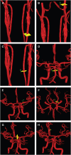
Measurement of volume flow rates in major cerebral vessels can be used to evaluate the hemodynamic effects of cerebrovascular disease. However, both age and vascular anatomy can affect flow rates independent of disease. We prospectively evaluated 325 healthy adult volunteers using phase contrast quantitative magnetic resonance angiography to characterize these effects on cerebral vessel flow rates and establish clinically useful normative reference values. Flows were measured in the major intracranial and extracranial vessels. The cohort ranged from 18 to 84 years old, with 157 (48%) females. All individual vessel flows and total cerebral blood flow (TCBF) declined with age, at 2.6 mL/minute per year for TCBF. Basilar artery (BA) flow was significantly decreased in individuals with one or both fetal posterior cerebral arteries (PCAs). Internal carotid artery flows were significantly higher with a fetal PCA and decreased with a hypoplastic anterior cerebral artery. Indexing vessel flows to TCBF neutralized the age effect, but anatomic variations continued to impact indexed flow in the BA and internal carotid artery. Variability in normative flow ranges were reduced in distal vessels and by examining regional flows. Cerebral vessel flows are affected by age and cerebrovascular anatomy, which has important implications for interpretation of flows in the disease state.
Figures


References
-
- Derdeyn CP, Grubb RL, Jr, Powers WJ. Cerebral hemodynamic impairment: methods of measurement and association with stroke risk. Neurology. 1999;53:251–259. - PubMed
-
- Amin-Hanjani S, Du X, Zhao M, Walsh K, Malisch TW, Charbel FT. Use of quantitative magnetic resonance angiography to stratify stroke risk in symptomatic vertebrobasilar disease. Stroke. 2005;36:1140–1145. - PubMed
-
- Amin-Hanjani S, Shin JH, Zhao M, Du X, Charbel FT. Evaluation of extracranial-intracranial bypass using quantitative magnetic resonance angiography. J Neurosurg. 2007;106:291–298. - PubMed
-
- Hendrikse J, van der Zwan A, Ramos LM, Tulleken CA, van der Grond J. Hemodynamic compensation via an excimer laser-assisted, high-flow bypass before and after therapeutic occlusion of the internal carotid artery. Neurosurgery. 2003;53:858–863. - PubMed
-
- Hendrikse J, Rutgers DR, Klijn CJ, Eikelboom BC, van der Grond J. Effect of carotid endarterectomy on primary collateral blood flow in patients with severe carotid artery lesions. Stroke. 2003;34:1650–1654. - PubMed
Publication types
MeSH terms
LinkOut - more resources
Full Text Sources
Other Literature Sources
Medical

