. 2015 Feb;35(2):221-9.
doi: 10.1038/jcbfm.2014.186. Epub 2014 Nov 12.

Sex differences in ischemic stroke sensitivity are influenced by gonadal hormones, not by sex chromosome complement

Affiliations

Sex differences in ischemic stroke sensitivity are influenced by gonadal hormones, not by sex chromosome complement

Bharti Manwani et al. J Cereb Blood Flow Metab. 2015 Feb.

Abstract

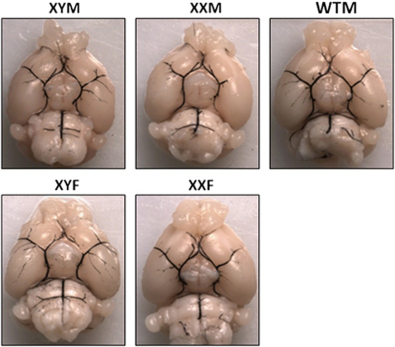
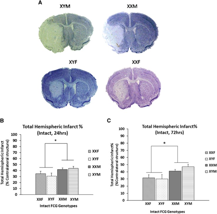
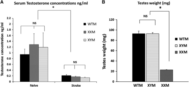
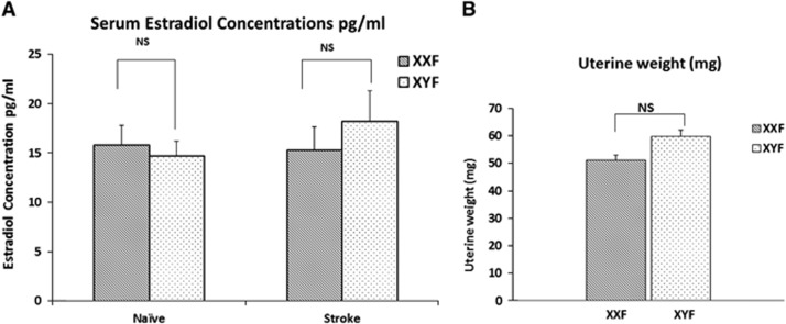
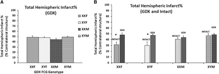
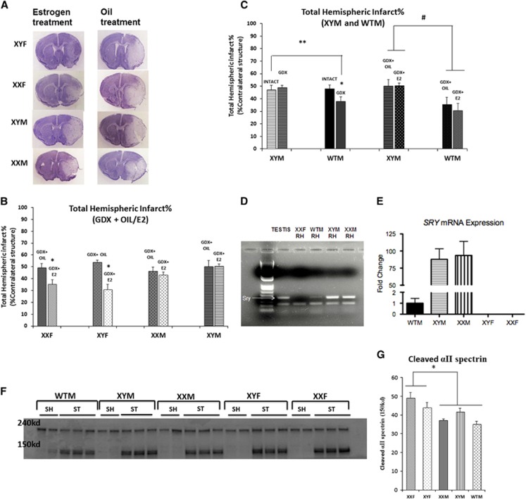
Epidemiologic studies have shown sex differences in ischemic stroke. The four core genotype (FCG) mouse model, in which the testes determining gene, Sry, has been moved from Y chromosome to an autosome, was used to dissociate the effects of sex hormones from sex chromosome in ischemic stroke outcome. Middle cerebral artery occlusion (MCAO) in gonad intact FCG mice revealed that gonadal males (XXM and XYM) had significantly higher infarct volumes as compared with gonadal females (XXF and XYF). Serum testosterone levels were equivalent in adult XXM and XYM, as was serum estrogen in XXF and XYF mice. To remove the effects of gonadal hormones, gonadectomized FCG mice were subjected to MCAO. Gonadectomy significantly increased infarct volumes in females, while no change was seen in gonadectomized males, indicating that estrogen loss increases ischemic sensitivity. Estradiol supplementation in gonadectomized FCG mice rescued this phenotype. Interestingly, FCG male mice were less sensitive to effects of hormones. This may be due to enhanced expression of the transgene Sry in brains of FCG male mice. Sex differences in ischemic stroke sensitivity appear to be shaped by organizational and activational effects of sex hormones, rather than sex chromosomal complement.


Figures

Figure 1
Figure 1
Vascular anatomy of large cerebral vessels in four core genotype (FCG) and wild-type male (WTM) control mice. No large vessel abnormalities are seen on gross inspection. There was no significant difference in PcomA plasticity score across the genotypes.
Figure 2
Figure 2
(A) Representative cresyl violet stained sections of the brains (90 minutes middle cerebral artery occlusion (MCAO), 24 hours end point) in the FCG mice. Significantly higher injury is seen in males as compared with females. (B) Total hemispheric infarct percentage at 24 hours end point after 90 minutes of MCAO. *Significant main effect of sex was seen. (C) Total hemispheric infarct percentage at 72 hours end point after 90 minutes of MCAO. *Significant main effect of sex was seen. FCG, four core genotype.
Figure 3
Figure 3
(A) Serum testosterone concentrations (ng/mL) as measured by enzyme-linked immunoassay (ELISA). There was no significant difference in the serum testosterone concentrations in XY male (XYM), XX male (XXM), and wild-type male (WTM) mice. Testosterone levels decreased significantly after middle cerebral artery occlusion (MCAO) (*significant stroke effect). There was no significant effect of the location of Sry (endogenous in WTM versus autosomal in XXM and XYM) or sex chromosome complement (XX/XY) on the serum testosterone levels. (B) Testicular weights (mg) in male FCG mice. There was no significant difference in the testicular weights of WTM's and XYM's (indicating that Sry transgene has no effect in testis weight). *Testicular weights of XXM's and XYM's were significantly different, P<0.05.
Figure 4
Figure 4
(A) Serum estradiol concentrations (pg/mL) as measured by enzyme-linked immunoassay (ELISA). There was no significant difference in the estradiol concentrations of XY female (XYF) and XX female (XXF) in the naïve or middle cerebral artery occlusion (MCAO) group. No stroke (MCAO) or sex chromosome complement (XX/XY) effect was seen. (B) Uterine weights (mg) in female FCG mice. There was no significant difference in the uterine weights of XYF and XXF mice.
Figure 5
Figure 5
(A) Total hemispheric infarct % in gonadectomized (GDX) four core genotype (FCG) mice. Infarct volumes at 72 hours after middle cerebral artery occlusion (MCAO) in the gonadectomized state. No significant effect of sex or sex chromosome complement was seen. (B) Total hemispheric infarct % in GDX versus Intact FCG mice. *Significant increase in infarct volumes after gonadectomy in female FCG mice.
Figure 6
Figure 6
(A) Cresyl violet sections of four core genotype (FCG) mice supplemented with oil/estrogen (E2) and subjected to middle cerebral artery occlusion (MCAO). (B) Total hemispheric infarct % in gonadectomized FCG mice supplemented with oil/estrogen (E2). *Female FCG mice have significantly lower infarct volumes in estrogen group as compared with oil group. (C) Total hemispheric infarct % in wild-type male (WTM) and XY male (XYM). *Significant decrease in infarct volumes was seen in GDX WTM mice versus Intact WTM. **Significant decrease in infarct volumes was seen in GDX WTM mice versus GDX/Intact XYM mice. # significant effect of Sry. (D) PCR showing Sry expression in the brain (RH, right hemisphere) of FCG and WTM mice. First two on the left are controls—testis sample from XYM mice (positive control), RH from female (XXF) (negative control). Higher expression of Sry is seen in the brains of FCG males—XYM and XX male (XXM) versus WTM. (E) Sry mRNA expression in naïve FCG and WTM mice. *Significant 90-fold increase in mRNA expression of Sry in the phenotypes with the exogenous Sry (XYM and XXM) compared with WTM (P<0.05). *No expression was seen in phenotypically female mice (XY female (XYF) and XX female (XXF)). (F, G) Western blot showing caspase mediated αII spectrin cleavage (150 kd) increased in gonadectomized females (XXF and XYF). The 240- kd band is the total αII spectrin. ST—stroke (MCAO); SH—sham mice. Graph in (G) shows a significant sex effect in cleaved αII spectrin expression, n=2 shams, n=3 strokes in each group. GDX, gonadectomy.

References

    1. Go AS, Mozaffarian D, Roger VL, Benjamin EJ, Berry JD, Borden WB, et al. Heart disease and stroke statistics—2013 update: A report from the american heart association. Circulation. 2013;127:e6–e245. - PMC - PubMed
    1. Hurn PD, Macrae IM. Estrogen as a neuroprotectant in stroke. J Cereb Blood Flow Metab. 2000;20:631–652. - PubMed
    1. Alkayed NJ, Harukuni I, Kimes AS, London ED, Traystman RJ, Hurn PD.Gender-linked brain injury in experimental stroke Stroke 199829159–165.discussion 166. - PubMed
    1. Yamori Y, Horie R, Sato M, Ohta K. Proceedings: Prophylactic trials for stroke in stroke-prone shr: Effect of sex hormones. Jpn Heart J. 1976;17:404–406. - PubMed
    1. Vannucci SJ, Hurn PD. Gender differences in pediatric stroke: is elevated testosterone a risk factor for boys. Ann Neurol. 2009;66:713–714. - PMC - PubMed

Publication types

MeSH terms