. 2015 Feb;19(2):408-17.
doi: 10.1111/jcmm.12461. Epub 2014 Nov 11.

Role of p38 MAPK in enhanced human cancer cells killing by the combination of aspirin and ABT-737

Affiliations

Role of p38 MAPK in enhanced human cancer cells killing by the combination of aspirin and ABT-737

Chong Zhang et al. J Cell Mol Med. 2015 Feb.

Abstract

Regular use of aspirin after diagnosis is associated with longer survival among patients with mutated-PIK3CA colorectal cancer, but not among patients with wild-type PIK3CA cancer. In this study, we showed that clinically achievable concentrations of aspirin and ABT-737 in combination could induce a synergistic growth arrest in several human PIK3CA wild-type cancer cells. In addition, our results also demonstrated that long-term combination treatment with aspirin and ABT-737 could synergistically induce apoptosis both in A549 and H1299 cells. In the meanwhile, short-term aspirin plus ABT-737 combination treatment induced a greater autophagic response than did either drug alone and the combination-induced autophagy switched from a cytoprotective signal to a death-promoting signal. Furthermore, we showed that p38 acted as a switch between two different types of cell death (autophagy and apoptosis) induced by aspirin plus ABT-737. Moreover, the increased anti-cancer efficacy of aspirin combined with ABT-737 was further validated in a human lung cancer A549 xenograft model. We hope that this synergy may contribute to failure of aspirin cancer therapy and ultimately lead to efficacious regimens for cancer therapy.

Keywords: ABT-737; aspirin; combination; p38.


Figures

Fig 1
Fig 1
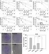
Combination cytotoxicity of aspirin and ABT-737. (A) The cells were incubated with the compounds for 72 hrs. Dose–response curves of human cancer cell lines to aspirin, ABT-737 or the combination were showed. Each condition had six replicates and error bars represented standard deviation. (B) Combination treatment with aspirin and ABT-737 inhibited the colony formation in A549 cells. Cells were treated with aspirin (1.25 mM), ABT-737 (2.5 μM) or the combination for 14 days. (C) Changes in the number of colonies formed by A549 cells after treatments. **P < 0.01, mono-treatment versus combination treatment.
Fig 2
Fig 2
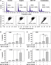
Long-term combination treatment with aspirin and ABT-737 caused enhanced apoptosis. (A) A549 cells were treated with aspirin (2.5 mM), ABT-737 (5 μM) or the combination for 48 hrs. Cells were stained with PI (upper) or JC-1 (bottom) and analysed by flow cytometry. A549 (B) and H1299 (C) cells in six-well plates were exposed to the compounds for 48 hrs and then cells were analysed by flow cytometry after PI staining. A549 (D) and H1299 (E) cells in six-well plates were exposed to compounds for 48 hrs and then cells were analysed by flow cytometry after JC-1 staining. The experiments were repeated three times and error bars represented standard deviation. *P < 0.05, **P < 0.01.
Fig 3
Fig 3
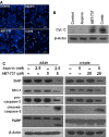
Long-term combination treatment with aspirin and ABT-737 caused activation of various apoptosis related proteins. (A) Aspirin plus ABT-737 induced apoptotic bodies in A549 cells. Nuclear DNA was visualized by DAPI staining; scale bar = 5 μm. (B) Cytochrome c release from mitochondria into cytosol was assessed at 48 hrs in A549 cells incubated with the agents. Cytosolic protein extracts were immunoblotted with specified antibodies for cytochrome c and β-actin. (C) A549 and H1299 cells were exposed to aspirin, ABT-737 or the combination for 48 hrs, after which protein extracts were immunoblotted with specified antibodies for XIAP, Mcl-1, caspase-3, cleaved caspase-3, PARP and β-actin.
Fig 4
Fig 4
Short-term combination treatment with aspirin and ABT-737-induced autophagy. (A) A549 cells were exposed to aspirin (2.5 mM), ABT-737 (5 μM) or the combination for 12 hrs, after which protein extracts were immunoblotted using LC-3. (B) LC-3 II/I ratios were significantly increased in combination treatment with aspirin (2.5 mM) plus ABT-737 (5 μM) for 12 hrs in A549 cells. (C) A549 cells were transfected with GFP-LC-3 plasmid according to manufacturer's recommendations. Twenty-four hours after transfection, cells were exposed to aspirin (2.5 mM), ABT-737 (5 μM) or the combination for 12 hrs. Aspirin plus ABT-737 induced GFP-LC3 dot formation in A549 cells; scale bar = 5 μm. (D) Quantified results of punctate GFP-LC3/A549 cells undergoing aspirin and/or ABT-737 treatment were measured (mean ± SD, n = 3). ***P < 0.001, **P < 0.01. A549 (E) and H1299 (F) cells treated with compounds for 12 hrs were stained with AO and analysed by flow cytometry. (G) A549 and H1299 cells were pre-treated with 1 mM 3-MA for 1 hr and incubated with aspirin and ABT-737 at the indicated concentrations for 12 hrs, after which protein extracts were immunoblotted using LC-3. (H) A549 and H1299 cells were pre-treated with 1 nM Baflomycine A1 for 1 hr and incubated with aspirin and ABT-737 at the indicated concentrations for 12 hrs, after which protein extracts were immunoblotted using LC-3. A549 (I) and H1299 (J) cells were pre-treated with 1 mM 3-MA for 1 hr and incubated with aspirin and/or ABT-737 at the indicated concentrations for 48 hrs. Percentages of apoptotic cells were determined by PI analysis. A549 (K) and H1299 (L) cells were pre-treated with 1 nM Baflomycine A1 for 1 hr and incubated with aspirin and/or ABT-737 at the indicated concentrations for 48 hrs. Percentages of apoptotic cells were determined by PI analysis.
Fig 5
Fig 5
P38 MAPK acted as a switch for the transition from autophagy to apoptosis during combination treatment with aspirin and ABT-737. (A) A549 cells were exposed to aspirin (2.5 mM), ABT-737 (5 μM) or the combination for 12 and 48 hrs, after which protein extracts were immunoblotted using p38, p-p38, LC-3, PARP and β-actin. (B) A549 cells were exposed to aspirin (2.5 mM), ABT-737 (5 μM) or the combination for 6, 12, 24 and 48 hrs, H1299 cells were exposed to aspirin (5 mM), ABT-737 (20 μM) or the combination for 6, 12, 24 and 48 hrs, after which protein extracts were immunoblotted using p-p38 and β-actin. (C) A549 cells were pre-treated with p38 MAPK inhibitor SB-203580 (10 μM) for 1 hr and then treated with 2.5 mM aspirin and/or 5 μM ABT-737 for 12 hrs, after which protein extracts were immunoblotted using p-p38, LC-3 and β-actin. (D) A549 cells were transfected with control SiRNA, p38 siRNA-1 and p38 siRNA-2 according to the manufacturer's recommendations. Forty-eight hours after transfection, cell lysates were prepared for Western blot analysis. (E) The expression of caspase-3 in A549 cells that had been transfected with p38 siRNA and treated with 2.5 mM aspirin, either alone or in combination with 5 μM ABT-737, for 48 hrs were examined.
Fig 6
Fig 6
The synergistic effect of aspirin and ABT-737 in a human lung cancer A549 xenograft model. (A) The mice transplanted with A549 human xenografts were randomly divided into four groups and given injection of aspirin (100 mg/kg, i.g.), ABT-737 (50 mg/kg, i.p.), the combination or vehicle for a period of 29 days. Relative tumour volume were expressed as mean ± SD (n = 8 per group). **Relative to control, P < 0.01; Relative to aspirin group, P < 0.05; §Relative to ABT-737 group, P < 0.05. (B) The average bodyweight of each group was expressed as mean ± SD (n = 8 per group).

References

    1. Coxhead JM, Williams EA, Mathers JC. DNA mismatch repair status may influence anti-neoplastic effects of butyrate. Biochem Soc Trans. 2005;33:728–9. - PubMed
    1. Pennarun B, Kleibeuker JH, Boersma-van Ek W, et al. Targeting FLIP and Mcl-1 using a combination of aspirin and sorafenib sensitizes colon cancer cells to TRAIL. J Pathol. 2013;229:410–21. - PubMed
    1. Feskanich D, Bain C, Chan AT, et al. Aspirin and lung cancer risk in a cohort study of women: dosage, duration and latency. Br J Cancer. 2007;97:1295–9. - PMC - PubMed
    1. Menezes RJ, Huber KR, Mahoney MC, et al. Regular use of aspirin and pancreatic cancer risk. BMC Public Health. 2002;2:18. - PMC - PubMed
    1. Printz C. Aspirin extends life of some patients with colorectal cancer. Cancer. 2013;119:472–3. - PubMed

Publication types

MeSH terms