

Editorial
doi: 10.1186/s12968-014-0089-6.
Probing dynamic myocardial microstructure with cardiac magnetic resonance diffusion tensor imaging
Affiliations
- PMID: 25388937
- PMCID: PMC4229597
- DOI: 10.1186/s12968-014-0089-6
Item in Clipboard
Editorial
Probing dynamic myocardial microstructure with cardiac magnetic resonance diffusion tensor imaging
J Cardiovasc Magn Reson.
.
Abstract
This article is an invited editorial comment on the paper entitled "In vivo cardiovascular magnetic resonance diffusion tensor imaging shows evidence of abnormal myocardial laminar orientations and mobility in hypertrophic cardiomyopathy" by Ferreira et al., and published as Journal of Cardiovascular Magnetic Resonance 2014; 16:87.
Figures
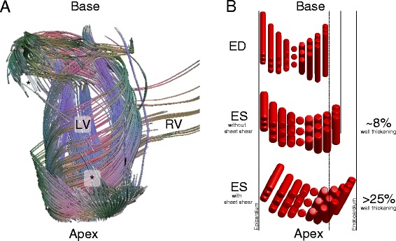
Myocardial structure and function.
(A) Diffusion tensor “fiber” tracking of a canine heart reveals large-scale connectivity of the end-to-end anastomoses of continuously branching myocytes. With sufficiently high spatial resolution (especially required at the apex and base) the principal eigenvector can be tracked from asterisk (*) to asterisk (*) while tracing out aspects of the base, apex, endocardium, and epicardium. (B) A representative sheetlet structure comprised of three myocyte layers. Incompressible myocyte shortening of ~15% gives rise to only ~8% radial wall thickening when sheet-shear is absent. In the presence of sheet shear, which is accommodated by the sheetlets, radial wall thickening increases to >25%.
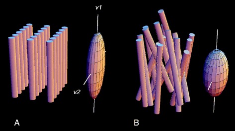
Schematic representation of architecture of fibers and sheets of normal myocardium (A) and in myocardial fiber disarray (B), and of their respective diffusion tensors. In the normal, fibers are locally parallel, and organized in local planes, sheetlets. Diffusion tensors represent an ellipsoid whose leading eigenvector (v1), the direction of maximum diffusion, is aligned with the mean axis of the myocardial fibers, and whose second eigenvector (v2) indicates the local sheet orientation. These directions are well-defined in the normal heart (A), and their distinctiveness is reduced or lost in fiber disarray. This is reflected in the lengths of the eigenvectors, which become more nearly equal in disarray (B). In maximum disorder, the diffusion tensor is a sphere, without distinguished directions. Details omitted include the facts that myocardial fibers are not isolated but branched syncytia, and that disarray is typically accompanied by increased matrix protein and non-contractile cells.
Comment on
-
In vivo cardiovascular magnetic resonance diffusion tensor imaging shows evidence of abnormal myocardial laminar orientations and mobility in hypertrophic cardiomyopathy.J Cardiovasc Magn Reson. 2014 Nov 12;16(1):87. doi: 10.1186/s12968-014-0087-8. J Cardiovasc Magn Reson. 2014. PMID: 25388867 Free PMC article.
References
-
- Streeter DD, Bassett DL. An engineering analysis of myocardial fiber orientation in pigs left ventricle in systole. Anat Rec. 1966;155(4):503. doi: 10.1002/ar.1091550403. - DOI
-
- Ferreira PF KP, McGill LA, Nielles-Vallespin S, Scott AD, Ho SY, McCarthy KP, Haba MM, Ismail TF, Gatehouse PD, de Silva R, Lyon AR, Prasad SK, Firmin DN, Pennell DJ. In vivo cardiovascular magnetic resonance diffusion tensor imaging shows evidence of abnormal myocardial laminar orientations and mobillity in hypertrophic cardiomyopathy. J Cardiovasc Magn Reson. 2014;16:87. doi: 10.1186/1532-429X-16-36. - DOI - PMC - PubMed
-
- LeGrice IJ, Smaill BH, Chai LZ, Edgar SG, Gavin JB, Hunter PJ. Laminar structure of the heart: ventricular myocyte arrangement and connective tissue architecture in the dog. Am J Physiol. 1995;269(2 Pt 2):H571–82. - PubMed
Publication types
MeSH terms
LinkOut - more resources
Full Text Sources
Other Literature Sources
