. 2015 Feb;19(2):463-73.
doi: 10.1111/jcmm.12478. Epub 2014 Nov 11.

Epidermal growth factor attenuates tubular necrosis following mercuric chloride damage by regeneration of indigenous, not bone marrow-derived cells

Affiliations

Epidermal growth factor attenuates tubular necrosis following mercuric chloride damage by regeneration of indigenous, not bone marrow-derived cells

Tzung-Hai Yen et al. J Cell Mol Med. 2015 Feb.

Abstract

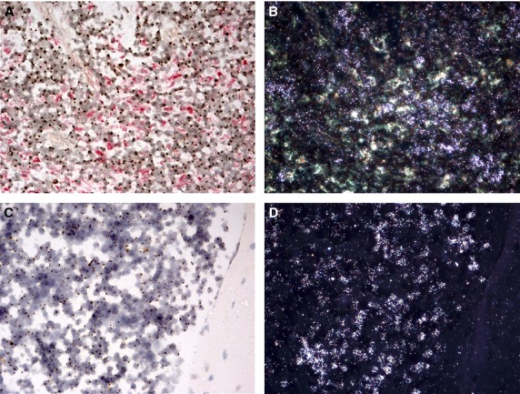
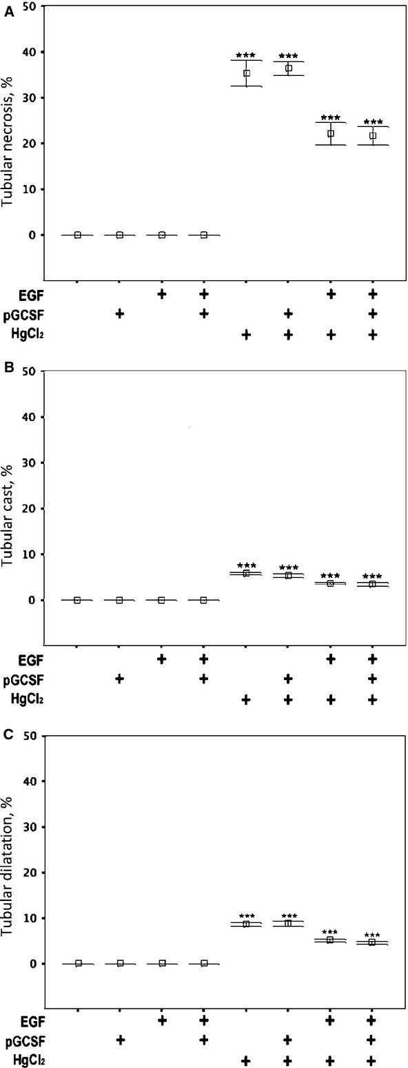
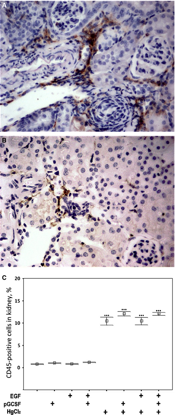
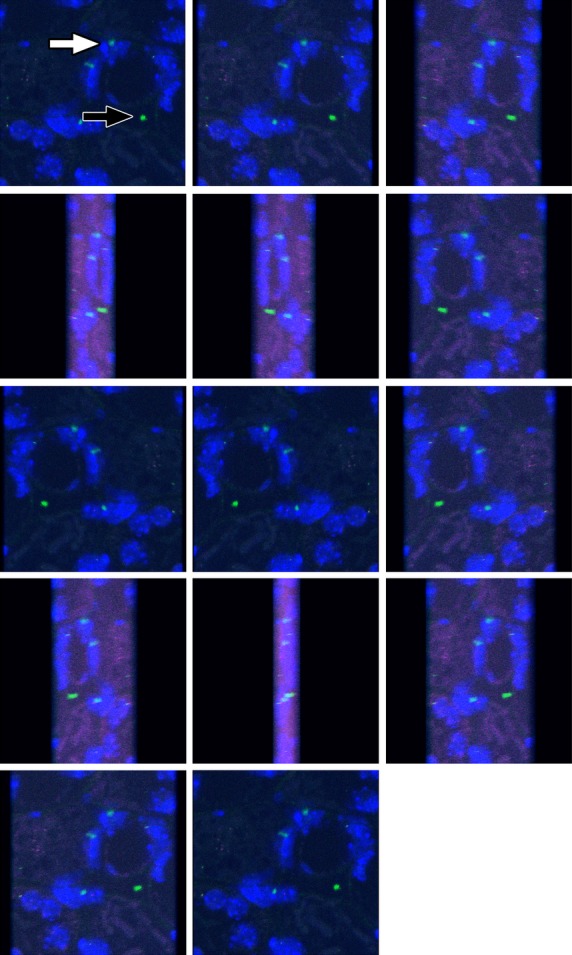
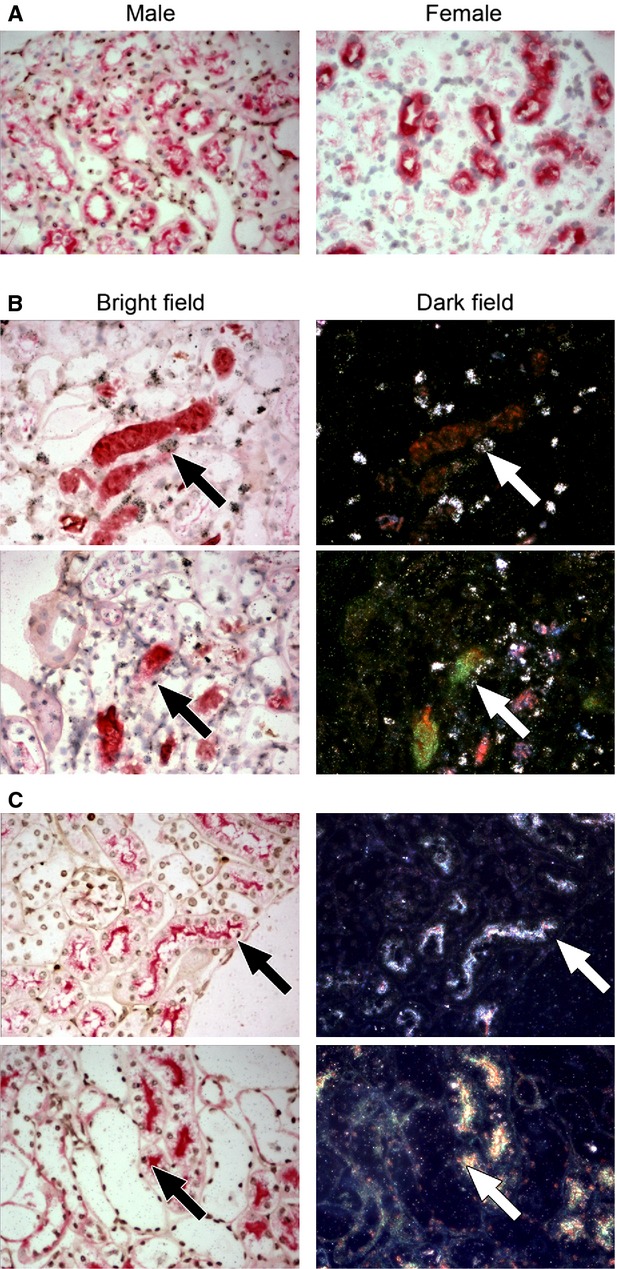
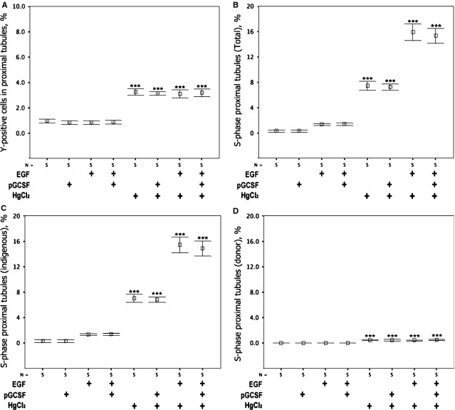
To assess effects of epidermal growth factor (EGF) and pegylated granulocyte colony-stimulating factor (P-GCSF; pegfilgrastim) administration on the cellular origin of renal tubular epithelium regenerating after acute kidney injury initiated by mercuric chloride (HgCl2 ). Female mice were irradiated and male whole bone marrow (BM) was transplanted into them. Six weeks later recipient mice were assigned to one of eight groups: control, P-GCSF+, EGF+, P-GCSF+EGF+, HgCl2 , HgCl2 +P-GCSF+, HgCl2 +EGF+ and HgCl2 +P-GCSF+EGF+. Following HgCl2 , injection tubular injury scores increased and serum urea nitrogen levels reached uraemia after 3 days, but EGF-treated groups were resistant to this acute kidney injury. A four-in-one analytical technique for identification of cellular origin, tubular phenotype, basement membrane and S-phase status revealed that BM contributed 1% of proximal tubular epithelium in undamaged kidneys and 3% after HgCl2 damage, with no effects of exogenous EGF or P-GCSF. Only 0.5% proximal tubular cells were seen in S-phase in the undamaged group kidneys; this increased to 7-8% after HgCl2 damage and to 15% after addition of EGF. Most of the regenerating tubular epithelium originated from the indigenous pool. BM contributed up to 6.6% of the proximal tubular cells in S-phase after HgCl2 damage, but only to 3.3% after additional EGF. EGF administration attenuated tubular necrosis following HgCl2 damage, and the major cause of this protective effect was division of indigenous cells, whereas BM-derived cells were less responsive. P-GCSF did not influence damage or regeneration.

Keywords: acute tubular necrosis; bone marrow-derived cells; epidermal growth factor; mercuric chloride; pegfilgrastim; tubular regeneration.


Figures

Fig 1
Fig 1
Confirmation of short-term haematopoietic reconstitution. Abundant Y-positive cells (brown nuclear dot) in the spleen (A and B) and BM (C and D) of a female recipient mouse indicate successful short-term haematopoietic reconstitution (bright field, A and C). Some of these Y-positive cells had incorporated 3H thymidine, detected by autoradiography as reflective silver grains under dark-field illumination (dark field, B and D).
Fig 2
Fig 2
Attenuation of acute kidney injury by EGF and the absence of effects of P-GCSF. (A) Prevalence of tubular cell necrosis, (B) cast formation and (C) tubular dilatation after various treatments.
Fig 3
Fig 3
Renal infiltration of leucocytes and macrophages after kidney damage and EGF and P-GCSF treatments. Immunohistochemistry for (A) CD45 and (B) F4/80 reveals clusters of infiltrated leucocytes and macrophages respectively 4 days after induction of acute kidney injury in a female recipient of male BM. This mouse was treated with HgCl2 and EGF, but without P-GCSF treatment. (C) In mice with renal damage, P-GCSF increased the abundance of CD45-positive cells in the kidney (10.46 ± 0.44% versus 0.8 ± 0.04%, P < 0.001). EGF injections did not affect the abundance of CD45-positive cells.
Fig 4
Fig 4
Four channel laser scanning confocal microscopy of a kidney section from a female mouse recipient of male bone marrow (BM), following mercuric chloride HgCl2 damage and EGF treatment. A gallery of images was generated from a projected Z-series to show how authentic fluorescence in situ hybridization signals for Y chromosomes (green) are present within cell nuclei (blue) (white arrow), whereas occasional green signals are artefactual (black arrow). The location of some BM-derived cells within tubular basement membranes is apparent from tissue architecture revealed by autofluorescence that has been captured in red and far-red channels. Authentic Y signals are always within the territory of a DAPI-stained nucleus.
Fig 5
Fig 5
Detection of bone marrow (BM)-derived tubular epithelium in S-phase using the ‘four-in-one’ technique. (A) Control tissues stained with biotinylated Phaseolus vulgaris leucoagglutinin lectin (red) and indirect in situ hybridization for Y chromosomes (brown) showing co-localization of staining in the proximal tubule in male proximal tubules and no Y signals in female tissue. (B and C) Regions of kidney from female recipients of male BM that received HgCl2 damage and EGF treatment stained using the four-in-one protocol; these examples show BM-derived proximal tubule epithelial cells either (B) with or (C) without incorporation of 3H thymidine that reveals the BM-derived epithelial cell to be in S-phase. The dark-field panels allow autoradiographic silver grains to be seen as bright white dots.
Fig 6
Fig 6
The effects of acute kidney injury, EGF and P-GCSF on the proportion of proximal tubular cell proliferation of BM-origin and on S-Phase status. (A) Acute kidney injury increased the proportion of proximal tubule cells derived from BM irrespective of treatment with EGF and/or P-GCSF, thus scoring of these cells was not affected by the influx of CD45 cells after P-GCSF. (B) The proportion of proximal tubule cells in S-phase was increased by acute kidney injury and EGF synergistically, whereas P-GCSF was without effect. (C) Indigenous proximal tubule cells responded to acute kidney injury and more so in combination with EGF, whereas P-GCSF had no effect on S-phase status. (D) A small but statistically significant increase occurred in the proportion of exogenous (BM-derived) proximal tubule cells in S-phase, with no effect of EGF or P-GCSF. Differences from corresponding control groups: ***P < 0.001. Means ± SEM, n = 5.

References

    1. Humes HD, Cieslinski DA, Coimbra TM, et al. Epidermal growth factor enhances renal tubule cell regeneration and repair and accelerates the recovery of renal function in postischemic acute renal failure. J Clin Invest. 1989;84:1757–61. - PMC - PubMed
    1. Norman J, Tsau YK, Bacay A, et al. Epidermal growth factor accelerates functional recovery from ischaemic acute tubular necrosis in the rat: role of the epidermal growth factor receptor. Clin Sci (Lond) 1990;78:445–50. - PubMed
    1. Coimbra TM, Cieslinski DA, Humes HD. Epidermal growth factor accelerates renal repair in mercuric chloride nephrotoxicity. Am J Physiol. 1990;259:F438–43. - PubMed
    1. Morin NJ, Laurent G, Nonclercq D, et al. Epidermal growth factor accelerates renal tissue repair in a model of gentamicin nephrotoxicity in rats. Am J Physiol. 1992;263:F806–11. - PubMed
    1. Poulsom R, Forbes SJ, Hodivala-Dilke K, et al. Bone marrow contributes to renal parenchymal turnover and regeneration. J Pathol. 2001;195:229–35. - PubMed

Publication types