. 2014 Nov 11;5(6):e02165.
doi: 10.1128/mBio.02165-14.

QseC inhibitors as an antivirulence approach for Gram-negative pathogens

Affiliations

QseC inhibitors as an antivirulence approach for Gram-negative pathogens

Meredith M Curtis et al. mBio. .

Abstract

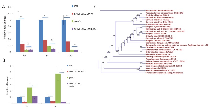
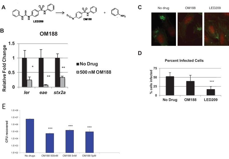
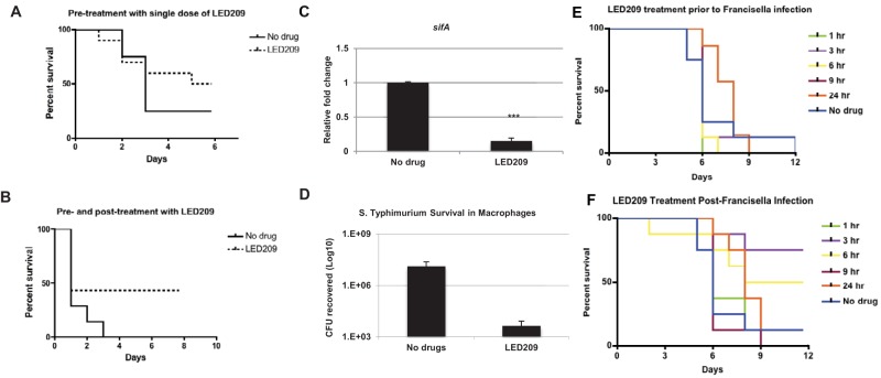
Invasive pathogens interface with the host and its resident microbiota through interkingdom signaling. The bacterial receptor QseC, which is a membrane-bound histidine sensor kinase, responds to the host stress hormones epinephrine and norepinephrine and the bacterial signal AI-3, integrating interkingdom signaling at the biochemical level. Importantly, the QseC signaling cascade is exploited by many bacterial pathogens to promote virulence. Here, we translated this basic science information into development of a potent small molecule inhibitor of QseC, LED209. Extensive structure activity relationship (SAR) studies revealed that LED209 is a potent prodrug that is highly selective for QseC. Its warhead allosterically modifies lysines in QseC, impairing its function and preventing the activation of the virulence program of several Gram-negative pathogens both in vitro and during murine infection. LED209 does not interfere with pathogen growth, possibly leading to a milder evolutionary pressure toward drug resistance. LED209 has desirable pharmacokinetics and does not present toxicity in vitro and in rodents. This is a unique antivirulence approach, with a proven broad-spectrum activity against multiple Gram-negative pathogens that cause mammalian infections.

Importance: There is an imminent need for development of novel treatments for infectious diseases, given that one of the biggest challenges to medicine in the foreseeable future is the emergence of microbial antibiotic resistance. Here, we devised a broad-spectrum antivirulence approach targeting a conserved histidine kinase, QseC, in several Gram-negative pathogens that promotes their virulence expression. The LED209 QseC inhibitor has a unique mode of action by acting as a prodrug scaffold to deliver a warhead that allosterically modifies QseC, impeding virulence in several Gram-negative pathogens.


Figures

FIG 1
FIG 1
LED209 targets the histidine sensor kinase QseC to attenuate virulence gene expression. (A, B) LED209 signals through the bacterial receptor QseC but not through the bacterial receptor QseE. Wild-type (WT) EHEC and the ΔqseC (A) and ΔqseE (B) isogenic mutants were grown in vitro in the presence (5 nM) or absence (DMSO only) of LED209 to late logarithmic phase. Expression of ler, the master transcriptional regulator of the LEE pathogenicity island in EHEC, of tir, a key LEE-encoded virulence gene, and of stx2a, a subunit of Shiga toxin, was measured by qRT-PCR. Expression is relative to that of the WT grown in the absence of LED209 (error bars, standard deviation [SD]; **, P < 0.01; ***, P < 0.001; NS, not significant). (C) QseC homologues are present in more than 25 plant and animal pathogens.
FIG 2
FIG 2
LED209 acts as a prodrug, cleaving an aniline group to expose its active component. (A) Upon interaction with QseC, LED209 breaks into the active component of LED209, OM188, and an aniline group. Schematic depicts the breakdown of LED209. OM188 contains an isothiocyanate (S=C=N) that is reactive to lysine and cysteine. (B) Treatment of EHEC with OM188 in vitro (by adding DMSO to the no-drug control and OM188 in DMSO at the beginning of the experiment) decreases expression of virulence genes. WT EHEC was grown in vitro in the presence (500 nM) or absence (DMSO only) of OM188 to late logarithmic phase. Expression of ler, eae, and stx2a was measured by qRT-PCR. Expression is relative to that of the WT grown in the absence of OM188 (error bars, SD; **, P < 0.01; *, P < 0.05). (C) Fluorescent actin staining assay of HeLa cells infected with EHEC grown in the absence (DMSO, no drug) or presence (5 nM) of OM188 or LED209, stained with fluorescein isothiocyanate-phalloidin (actin, green) and propidium iodide (bacterial and HeLa DNA, red). Original magnification, ×63. (D) Quantification of fluorescent actin staining assay of the percentage of HeLa cells infected, as defined by pedestal formation by EHEC (n = 100 cells). (E) Intramacrophage replication of S. enterica Typhimurium in the presence and absence of OM188 (error bars, SD; ***, P < 0.001; **, P < 0.01; *, P < 0.05).
FIG 3
FIG 3
LED209 targets the sensor kinase QseC on a conserved lysine residue. (A) Schematic of the “click chemistry” approach used to identify the conserved lysine residue to which QseC binds. The warhead of LED209, OM188, was engineered to contain an azide group that will “click” with a fluorophore alkyne-containing group. The “click” compound was named OM201. (B) OM201 binds to QseC in vitro. Membrane preparations of the ΔqseC mutant with empty vector (C [no QseC]) or expressing QseC (pVS155) were incubated with 100 µM OM201 and then run on an SDS-PAGE gel to separate proteins. (C) qRT-PCR of EHEC virulence genes (ler, tir, and stx2) in the absence or presence of OM201 (5 nM) showing that OM201 is active to inhibit virulence gene expression. (D) Two conserved lysine residues were identified through mass spectrometric analysis as candidates to covalently bind to OM188. Membrane preparations preincubated with OM188 were submitted for peptide analysis by LC-MS. Peptide fragments containing lysine 256 (K256) and lysine 427 (K427) were bound to OM188 and predicted to be sites of covalent attachment. (E) Alignment of QseC peptide sequences in EHEC, S. Typhimurium, and F. tularensis indicates that K256 is conserved in all three species. (F) Mutation of conserved lysines K256 and K427 result in a loss of virulence. qRT-PCR of the expression of the EHEC virulence genes: ler encodes a transcriptional activator of virulence genes, tir encodes a key virulence factor, and stx2 encodes the Shiga toxin in the ΔqseC (VS138) with qseC WT (pVS155), ΔqseC (VS138), ΔqseC (VS138) with the K256R or K427R qseC mutants. ***, P < 0.001.
FIG 4
FIG 4
Minor modifications to the structure of LED209 significantly impact its activity. (A) Four major components of LED209: the left-hand aniline group (part A), the thiourea (part B), the sulfur dioxide and amine group (part C), and the right-hand benzene (part D) were modified for structure analysis relationship (SAR) studies. Structures of the modified compounds are depicted in panel A. (B) Modifications to LED209 impact its ability to attenuate virulence gene expression in EHEC. WT EHEC was grown in vitro in the presence (5 nM) or absence (DMSO only) of modified compounds to late logarithmic phase. Expression of ler and stx2a was measured by qRT-PCR. Expression is relative to WT grown in the absence of drug (error bars, SD; *, P < 0.01). (C) Commercially available LED209 (Sigma) loses its efficacy with the addition of a hydrate at an undisclosed location. WT EHEC was grown in the presence (5 nM) of either LED209 synthesized at UT Southwestern or LED209 hydrate commercially available through Sigma-Aldrich, and expression of ler and stx2a was measured by qRT-PCR. Expression is relative to WT grown in the absence of drug (error bars, SD; ***, P < 0.001). (D) Fluorescent actin staining assay of HeLa cells infected with EHEC grown in the absence (DMSO, no drug) or presence (5 nM) of LED209 or LED209 hydrate (Sigma), stained with fluorescein isothiocyanate-phalloidin (actin, green) and propidium iodide (bacterial and HeLa DNA, red). Original magnification, ×63. (E) Quantification of fluorescent actin staining assay of the percentage of HeLa cells infected, as defined by pedestal formation by EHEC (n = 100 cells; error bars, SD; **, P < 0.01). (F) LED209 manufactured according to Good Laboratory Practice (GLP) standards was tested for its ability to inhibit EHEC virulence gene expression. WT EHEC was grown in the presence or absence of LED209 GLP, and expression of ler and stx2a was measured by qRT-PCR. Expression is relative to WT grown in the absence of drug (error bars, SD; ***, P < 0.001).
FIG 5
FIG 5
Treatment of LED209 protects against S. Typhimurium and F. tularensis. (A, B) Administration of LED209 protects BALB/c mice from S. Typhimurium infection. Mice were administered a single dose of LED209 (20 mg/kg) at 3 h before (A) or multiple doses of LED209 (20 mg/kg) at 3 h before, at the time of, and 3, 6, 9, and 12 h after (B) intraperitoneal injection of a lethal dose of S. Typhimurium (106 CFU). Graphs indicate survival to infection (n = 10). (C) Treatment of S. Typhimurium with 50 nM LED209 in vitro decreased the expression of sifA, an effector required for vacuolar replication. qRT-PCR of sifA in S. Typhimurium grown in N-minimal medium to late logarithmic phase. Expression is relative to S. Typhimurium grown in the absence of drug (error bars, SD; ***, P < 0.001). (D) J774 murine macrophages were infected with opsonized S. Typhimurium in the presence (5 nM) or absence (DMSO only) of LED209. Graph indicates intracellular survival within macrophages. (E, F) Administration of LED209 postinfection protects C3H/HeN mice from F. tularensis infection. Mice were administered a single dose of LED209 (20 mg/kg) at 1, 3, 6, 9, or 24 h prior to infection and then intranasally infected with 30 CFU of F. tularensis strain SCHU S4 (E) or a single dose at 1, 3, 6, 9, or 24 h postinfection with 30 CFU of F. tularensis (F). Graphs indicate survival to infection (n = 10).
FIG 6
FIG 6
LED209 diminishes biofilm formation in EAEC O104:H4 and in clinical isolates. (A) Treatment with LED209 reduced biofilm formation in the EAEC O104:H4 strain isolated from the 2011 German outbreak and in the UPEC strain UTI89. EAEC O104:H4, UTI89, and CFT073 were grown statically in LB in 96-well plates for 24 h in the presence (5 nM) or absence (DMSO only) of LED209. Biofilm formation was determined by measuring crystal violet binding at OD600 following gentle washing to remove unattached bacteria. (B) Treatment with LED209 reduced expression of stx2a, a subunit of Shiga toxin, in EAEC O104:H4. EAEC O104:H4 was grown in the presence (5 nM) or absence (DMSO) of LED209 to late logarithmic phase. Expression is relative to EAEC in the absence of drug. (C) Administration of LED209 diminished biofilm formation in clinical isolates isolated from urine and bladder biopsy samples. The isolates were grown statically in LB as described above, and biofilm formation was determined by measuring crystal violet binding at OD600 (error bars, SD; ***, P < 0.001; **, P < 0.01; *, P < 0.05). (D) Clinical samples were enriched in LB broth and isolated in MacConkey agar to differentiate Gram-negative bacteria. Isolates were classified via API 20E kits (bioMérieux, France). Probable species include Pseudomonas, Klebsiella, and Enterobacter species.

References

    1. Rasko DA, Sperandio V. 2010. Anti-virulence strategies to combat bacteria-mediated disease. Nat. Rev. Drug Discov. 9:117–128. 10.1038/nrd3013. - DOI - PubMed
    1. Petri WA, Jr, Miller M, Binder HJ, Levine MM, Dillingham R, Guerrant RL. 2008. Enteric infections, diarrhea, and their impact on function and development. J. Clin. Invest. 118:1277–1290. 10.1172/JCI34005. - DOI - PMC - PubMed
    1. Kostakioti M, Hadjifrangiskou M, Cusumano CK, Hannan TJ, Janetka JW, Hultgren SJ. 2012. Distinguishing the contribution of type 1 pili from that of other QseB-misregulated factors when QseC is absent during urinary tract infection. Infect. Immun. 80:2826–2834. 10.1128/IAI.00283-12. - DOI - PMC - PubMed
    1. Rasko DA, Moreira CG, Li de R, Reading NC, Ritchie JM, Waldor MK, Williams N, Taussig R, Wei S, Roth M, Hughes DT, Huntley JF, Fina MW, Falck JR, Sperandio V. 2008. Targeting QseC signaling and virulence for antibiotic development. Science 321:1078–1080. 10.1126/science.1160354. - DOI - PMC - PubMed
    1. Moreira CG, Weinshenker D, Sperandio V. 2010. QseC mediates Salmonella enterica serovar Typhimurium virulence in vitro and in vivo. Infect. Immun. 78:914–926. 10.1128/IAI.01038-09. - DOI - PMC - PubMed

Publication types

MeSH terms