. 2014 Nov 11;55(12):8163-72.
doi: 10.1167/iovs.14-14029.

Age- and race-related differences in human scleral material properties

Affiliations

Age- and race-related differences in human scleral material properties

Rafael Grytz et al. Invest Ophthalmol Vis Sci. .

Abstract

Purpose: We tested the hypothesis that there are age- and race-related differences in posterior scleral material properties, using eyes from human donors of European (20-90 years old, n = 40 eyes) and African (23-74 years old, n = 22 eyes) descent.

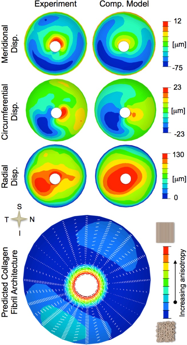
Methods: Inflation tests on posterior scleral shells were performed while full-field, three-dimensional displacements were recorded using laser speckle interferometry. Scleral material properties were fit to each eye using a microstructure-based constitutive formulation that incorporates the collagen fibril crimp and the local anisotropic collagen architecture. The effects of age and race were estimated using Generalized Estimating Equations, while accounting for intradonor correlations.

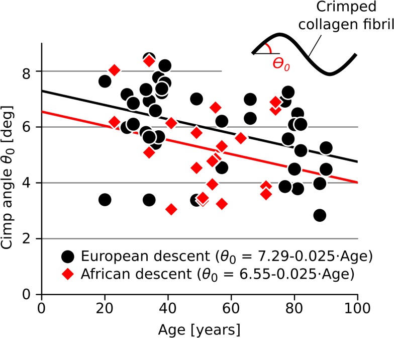
Results: The shear modulus significantly increased (P = 0.038) and collagen fibril crimp angle significantly decreased with age (P = 0.002). Donors of African descent exhibited a significantly higher shear modulus (P = 0.019) and showed evidence of a smaller collagen fibril crimp angle (P = 0.057) compared to donors of European descent. The in-plane strains in the peripapillary sclera were significantly lower with age (P < 0.015) and African ancestry (P < 0.015).

Conclusions: The age- and race-related differences in scleral material properties result in a loss of scleral compliance due to a higher shear stiffness and a lower level of stretch at which the collagen fibrils uncrimp. The loss of compliance should lead to larger high frequency IOP fluctuations and changes in the optic nerve head (ONH) biomechanical response in the elderly and in persons of African ancestry, and may contribute to the higher susceptibility to glaucoma in these at-risk populations.

Keywords: computational modeling; extracellular matrix; glaucoma posterior segment.


Figures

Figure 1
Figure 1
Tissue structures at multiple scales in the experiment (top) and computational model (bottom). Macro-level: The posterior scleral shell clamped into a pressurization apparatus and the eye-specific FE mesh. Meso-level: The collagen architecture showing a preferred circumferential alignment (arrow) represented by a von Mises distribution of collagen fibrils in the computer model. Micro-level: Crimped collagen fibrils in the sclera and in the computational model with the crimp angle θ0 shown.
Figure 2
Figure 2
Typical inverse numerical characterization of a scleral shell (left eye of a 77-year-old donor of African descent). Top: Comparison of experimentally measured displacements and the displacements predicted by the inverse computational model for an IOP elevation from 5 to 45 mm Hg. Bottom: Predicted anisotropic collagen architecture showing the predominant collagen fibril orientations (white lines) and the degree of anisotropy (contour plot). A distinct ring of circumferentially-aligned fibrils can be seen around the scleral canal, which was common to all eyes.
Figure 3
Figure 3
Fitted material parameters versus age plots. (A) Fitted shear modulus μ versus age, showing a significantly increasing shear modulus with age (P = 0.038) and a significantly higher shear modulus in donors of African descent (P = 0.019). (B) Fitted elastic modulus of collagen fibrils Efib versus age, showing a significant increase with age in the European (solid black line, P < 0.001) but not in the African descent group (dashed red line, P = 0.11). The aging trends in Efib also were significantly different between racial groups (P = 0.018). material parameters (μ, Efib) were expressed on a logarithmic scale for the statistical analysis.
Figure 4
Figure 4
Fitted crimp angle of collagen fibrils versus age. The fitted crimp angles show a significantly decreasing trend with age (P = 0.002) and evidence for a lower crimp angle in donors of African descent (P = 0.057).
Figure 5
Figure 5
Average scleral thickness versus age by racial group. There is no significant change in scleral thickness with age within either racial group (P > 0.05) and no significantly different trends with age between the racial groups (P > 0.05). The dashed lines represent the (nonsignificant) trend lines for the European (black) and African descent group (red).
Figure 6
Figure 6
Parameter study showing the in-plane strain response of the peripapillary sclera and using the full range of fitted material parameters found in this study. All plots were calculated using the same computer model (scleral shell with fitted material parameters that were closest to the mean values). The mean response (black line) was calculated using the mean fitted material parameters obtained in this study (μ = 0.35 MPa, Efib = 58.86 MPa, θ0 = 5.68°, R0/r0 = 5.3). The limit cases were calculated using the maximum (red dashed line with triangles up) and minimum (red dashed line with triangles down) values of each of the following material parameters in turn, while keeping the other material parameters at their mean values: (A) shear modulus μ, (B) elastic modulus of collagen fibrils Efib, and (C) crimp angle θ0. The in-plane strain was more sensitive to changes in the scleral shear modulus and crimp angle of the collagen fibrils compared to the elastic modulus of the collagen fibrils.
Figure 7
Figure 7
In-plane strain in the peripapillary sclera of donors younger (black line) and older than 50 years (blue line) versus IOP (mean, SD). In-plane strains are significantly lower in younger donors (P < 0.015). The age-related decrease in in-plane strain is mainly caused by the increasing scleral shear stiffness (μ) and the decreasing collagen fibril crimp (θ0) with age. Note that the arbitrary threshold of 50 years of age was only used to illustrate the age-related decrease in in-plane strain for Figure 5, while all statistical models assessed the effect of age as a continuous variable.
Figure 8
Figure 8
In-plane strain in the peripapillary sclera of donors of European descent (black line) and African descent (red line) versus IOP (mean, SD). In-plane strains are significantly lower in donors of African descent (P < 0.015). The race-related decrease in in-plane strain is mainly caused by a higher scleral shear stiffness (μ) and a lower collagen fibril crimp (θ0) in the African descent group.

References

    1. Nickells RW. The cell and molecular biology of glaucoma: mechanisms of retinal ganglion cell death. Invest Ophthalmol Vis Sci. 2012; 53: 2476–2481. - PMC - PubMed
    1. Nouri-Mahdavi K, Hoffman D, Coleman AL, et al. Predictive factors for glaucomatous visual field progression in the Advanced Glaucoma Intervention Study. Ophthalmology. 2004; 111: 1627–1635. - PubMed
    1. Leske MC, Heijl A, Hussein M, et al. Factors for glaucoma progression and the effect of treatment: the early manifest glaucoma trial. Arch Ophthalmol. 2003; 121: 48–56. - PubMed
    1. Gillespie BW, Musch DC, Guire KE, et al. The collaborative initial glaucoma treatment study: baseline visual field and test-retest variability. Invest Ophthalmol Vis Sci. 2003; 44: 2613–2620. - PubMed
    1. Drance S, Anderson DR, Schulzer M; Collaborative Normal-Tension Glaucoma Study Group. Risk factors for progression of visual field abnormalities in normal-tension glaucoma. Am J Ophthalmol. 2001; 131: 699–708. - PubMed

Publication types