. 2014 Nov 11;7(351):ra106.
doi: 10.1126/scisignal.2005375.

Inflammatory stimuli induce inhibitory S-nitrosylation of the deacetylase SIRT1 to increase acetylation and activation of p53 and p65

Affiliations

Inflammatory stimuli induce inhibitory S-nitrosylation of the deacetylase SIRT1 to increase acetylation and activation of p53 and p65

Shohei Shinozaki et al. Sci Signal. .

Abstract

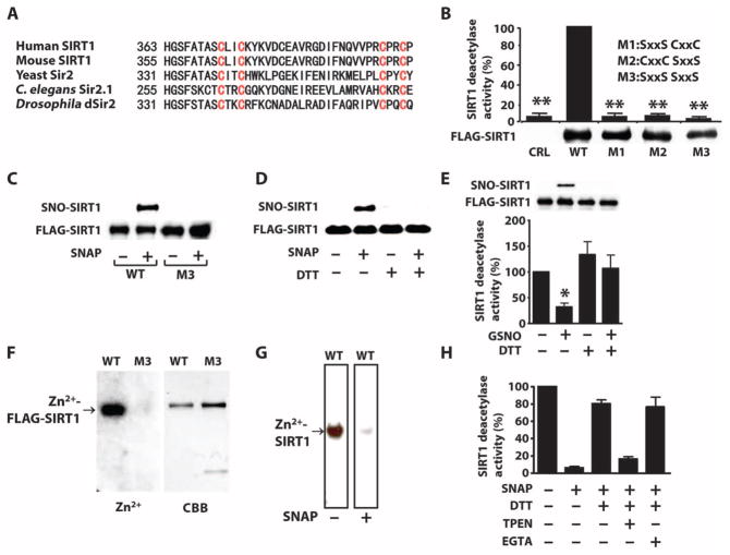
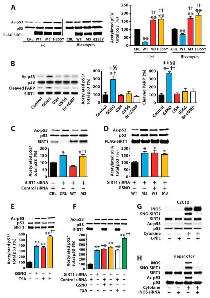
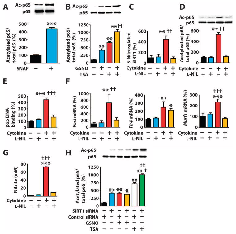
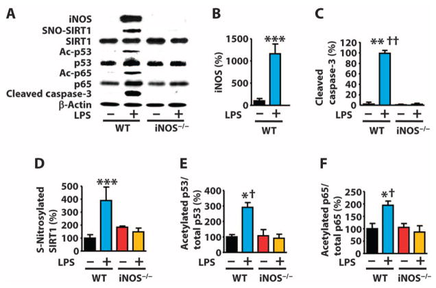
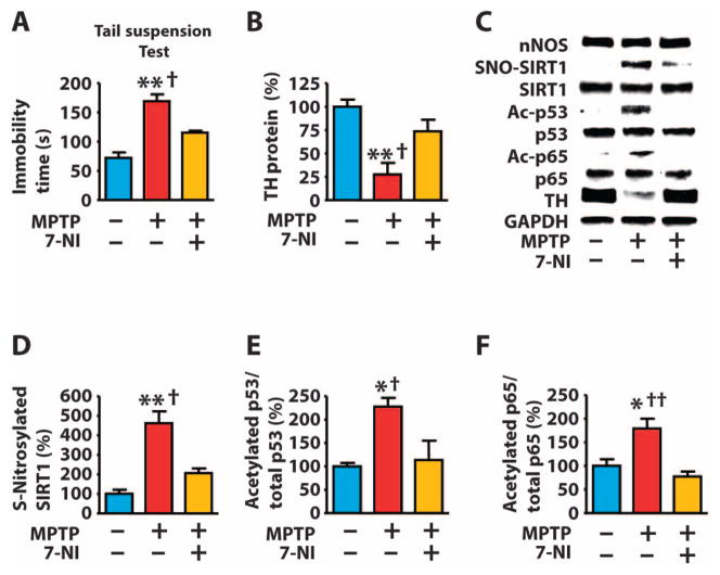
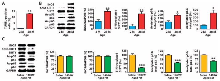
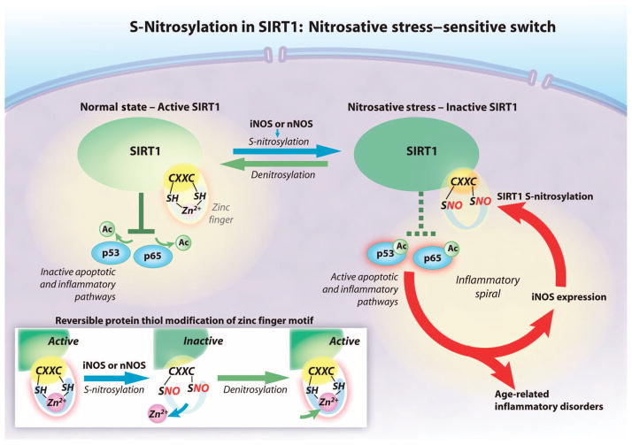
Inflammation increases the abundance of inducible nitric oxide synthase (iNOS), leading to enhanced production of nitric oxide (NO), which can modify proteins by S-nitrosylation. Enhanced NO production increases the activities of the transcription factors p53 and nuclear factor κB (NF-κB) in several models of disease-associated inflammation. S-nitrosylation inhibits the activity of the protein deacetylase SIRT1. SIRT1 limits apoptosis and inflammation by deacetylating p53 and p65 (also known as RelA), a subunit of NF-κB. We showed in multiple cultured mammalian cell lines that NO donors or inflammatory stimuli induced S-nitrosylation of SIRT1 within CXXC motifs, which inhibited SIRT1 by disrupting its ability to bind zinc. Inhibition of SIRT1 reduced deacetylation and promoted activation of p53 and p65, leading to apoptosis and increased expression of proinflammatory genes. In rodent models of systemic inflammation, Parkinson's disease, or aging-related muscular atrophy, S-nitrosylation of SIRT1 correlated with increased acetylation of p53 and p65 and activation of p53 and NF-κB target genes, suggesting that S-nitrosylation of SIRT1 may represent a proinflammatory switch common to many diseases and aging.


Conflict of interest statement

Competing interests: All authors declare that they have no competing interests. J. Stamler has licensed technology to Novartis within the field of S-nitrosylation that does not have a direct relationship to this work.

Figures

Fig. 1
Fig. 1. S-Nitrosylation of the CXXC motif reversibly inactivates SIRT1
(A) Amino acid sequence of the region containing the CXXC motifs in SIRT1 homologs. (B) Graph of the deacetylase activity of FLAG-wild-type (WT) or mutated SIRT1 (M1, M2, or M3) immunopurified from COS-7 cells. CRL, control plasmid. Western blot shows the relative abundance of these proteins in transfected cells. P < 0.01 CRL, M1, M2, or M3 compared to WT, Kruskal-Wallis test. Data are means ± SEM of three independent experiments. (C and D) Western blots for S-nitrosylated SIRT1 (SNO-SIRT1) or SIRT1 in lysates from COS-7 cells transfected FLAG-tagged WT SIRT1 or SIRT1M3 and exposed to the NO donor SNAP (300 μM) and carmustine (80 μM) (C) or recombinant SIRT1 incubated with SNAP or DTT (D). Data are representative of three independent experiments. (E and H) Graphs of the deacetylase activity of immunopurified FLAG-SIRT1 from COS-7 cells exposed to GSNO (0.6 mM) (E) or recombinant SIRT1 exposed to SNAP (0.6 mM) (H). Immunopurified FLAG-SIRT1 or recombinant proteins were incubated with the reducing agent DTT (5 mM), the Zn2+ chelator TPEN (200 μM), or the Ca2+ chelator EGTA (0.6 mM). Western blot in (E) shows the relative S-nitrosylation of SIRT1 used in this assay. *P < 0.05, one-way analysis of variance (ANOVA) with the Newman-Keuls post hoc test compared to control (E); P < 0.02 between samples, Kruskal-Wallis test (H). Data are means ± SEM of three independent experiments. (F and G) Autoradiographs of 65Zn2+ bound to immunopurified FLAG-SIRT1, but not FLAG-SIRT1M3 (F) or recombinant SIRT1 (G). In (G), 65Zn2+ binding is shown for the same membrane before and after incubation. Coomassie brilliant blue (CBB) shows total protein. Data are representative of two independent experiments.
Fig. 2
Fig. 2. SIRT1 S-nitrosylation mediates NO-induced p53 acetylation, leading to cell death
(A) Western blot of lysates from COS-7 cells transfected with WT or mutated SIRT1 and treated with bleomycin (10 μg/ml) or vehicle (dimethyl sulfoxide). CRL, control plasmid. **P < 0.01 compared to control plasmid; ††P < 0.01 compared to WT. One-way ANOVA with Tukey’s post hoc test. Data are means ± SEM of three biological replicates from one of three independent experiments. (B) Western blots of lysates from H1299 cells expressing exogenous wild-type exposed to GSNO (0.5 mM), reduced (GSH; 0.5 mM) or oxidized glutathione (GSSG; 0.5 mM), or 8-bromo cGMP (Br-cGMP; 0.5 mM). *P < 0.05, **P < 0.01 compared to control; P < 0.05, ††P < 0.01 compared to GSH; P < 0.05, ‡‡P < 0.01 compared to GSSG; §§P < 0.01 compared to Br-cGMP. One-way ANOVA with Tukey’s post hoc test. Data are means ± SEM from three independent experiments. (C to F) Western blot of lysates from COS-7 cells transfected with the indicated siRNAs and transfected with WT SIRT1, SIRT1M3, or control plasmids, or exposed to GSNO (1 mM) with or without TSA (1 μM). *P < 0.05 compared to control siRNA + control plasmid, ††P < 0.01 compared to SIRT1 siRNA + WT SIRT1 (C); *P < 0.05 compared to WT SIRT1 without GSNO (D); **P < 0.01 compared to control, ††P < 0.01 compared to TSA alone, ‡‡P < 0.01 compared to GSNO alone (E); **P < 0.01 compared to control siRNA alone, ††P < 0.01 compared to control siRNA + TSA (F). One-way ANOVA with Tukey’s post hoc test. Data are means ± SEM of three biological replicates from one of two independent experiments. (G and H) Western blot of C2C12 myotubes exposed to LPS (10μg/ml)andcytokines[TNF-α (10ng/ml)andIFN-γ (200ng/ml)]orHepa1c1c7 cells exposed to LPS (10 μg/ml) and cytokines [TNF-α (5 ng/ml) and IFN-γ (50 ng/ml)] with or without the iNOS inhibitor L-NIL (200 μM) or iNOS knockdown. Blots are representative of three independent experiments.
Fig. 3
Fig. 3. S-Nitrosylation–mediated inactivation of SIRT1 leads to increased acetylation of p65
(A and B) Western blots of lysates from COS-7 cells exposed to SNAP (300 μM) (A) or GSNO (1 mM) and TSA (1 μM) (B). (C to G) C2C12 myotubes exposed to LPS (10 μg/ml) and cytokines [TNF-α (10 ng/ml) and IFN-γ (200 ng/ml)] with or without L-NIL (200 μM). (C and D) Western blot. (E) p65 DNA binding activity. (F) mRNA abundance of NF-κB target genes. (G) Nitrite in the culture media. *P < 0.05, **P < 0.01, ***P < 0.001 compared to control; ††P < 0.01, †††P < 0.001 compared to cytokine + L-NIL. One-way ANOVA with Tukey’s post hoc test. (H) Western blot of lysates from COS-7 cells transfected with the indicated siRNAs and exposed to GSNO (1 mM) with or without TSA (1 μM). **P < 0.01 compared to control, P < 0.05 compared to control siRNA + TSA; ‡‡P < 0.01 compared to SIRT1 siRNA + GSNO. One-way ANOVA with Tukey’s post hoc test. Data are means ± SEM of three biological replicates from one of two (B and H) or three (A and C to G) independent experiments.
Fig. 4
Fig. 4. iNOS deficiency prevents endotoxin-induced SIRT1 S-nitrosylation and acetylation of p53 and p65 in mouse liver
(A to F) Western blot and quantification of lysates from livers of WT mice or iNOS knockout (iNOS−/−) mice injected with LPS [27.5 mg/kg, intraperitoneal (ip)]. *P < 0.05, **P < 0.01, ***P < 0.001 compared to WT without LPS; P < 0.05, †††P < 0.001 compared to iNOS−/− with LPS. One-way ANOVA with Tukey’s post hoc test. n = 8 mice per group.
Fig. 5
Fig. 5. S-Nitrosylation of SIRT1 parallels acetylation of p53 and p65 in a mouse model of Parkinson’s disease
(A to F) Mice were injected with MPTP (80 mg/kg, ip) and the nNOS inhibitor 7-NI (50 mg/kg, ip) and monitored 7 days after MPTP injection for bradykinesia (prolonged immobility time during the tail suspension test) (A), or the nigra-striatum was dissected 3 days after MPTP injection and analyzed by Western blot for the indicated proteins (B to F). *P < 0.05, **P < 0.05 compared to control; P < 0.05, ††P < 0.01 compared to MPTP + 7-NI. One-way ANOVA with Tukey’s post hoc test. n = 8 mice per group.
Fig. 6
Fig. 6. Increased abundance of iNOS is associated with S-nitrosylation of SIRT1 and acetylation and activation of p53 and p65 in skeletal muscle of aged rats
(A and B) Terminal deoxynucleotidyl transferase–mediated deoxyuridine triphosphate nick end labeling (TUNEL) (A) or Western blot of lysates (B) of skeletal muscle from rats at the indicated ages. *P < 0.05, **P < 0.01, 28 months (28 M) compared to 2 months (2 M). Unpaired two-tailed Student’s t test. n = 8 mice per group. (C) Western blot of skeletal muscle of 23-month-old rats injected with saline or the iNOS inhibitor 1400W (10 mg/kg, ip) for 10 days. **P < 0.01, ***P < 0.001 compared to saline. Unpaired two-tailed Student’s t test. n = 7 mice per group.
Fig. 7
Fig. 7. The proposed SIRT1 nitrosative stress–sensitive switch
SIRT1 decreases apoptosis and inflammation by deacetylating proteins in the p53 and NF-κB pathways. SIRT1 deacetylates p53 and the p65 subunit of NF-κB when the CXXC zinc finger motif is bound to zinc (Zn2+) (solid green). NO derived from iNOS or nNOS S-nitrosylates the cysteine thiol groups (SNO) in the CXXC motif, disrupting zinc binding, which renders SIRT1 inactive. Acetylation of p53 and p65 increases apoptosis and inflammation (dashed green). S-Nitrosylation and resultant inactivation of SIRT1 are reversible. Ac, acetyl group; SH, sulfhydryl group.

References

    1. Perreault M, Marette A. Targeted disruption of inducible nitric oxide synthase protects against obesity-linked insulin resistance in muscle. Nat Med. 2001;7:1138–1143. - PubMed
    1. Sugita H, Fujimoto M, Yasukawa T, Shimizu N, Sugita M, Yasuhara S, Martyn JA, Kaneki M. Inducible nitric-oxide synthase and NO donor induce insulin receptor substrate-1 degradation in skeletal muscle cells. J Biol Chem. 2005;280:14203–14211. - PubMed
    1. Cho DH, Nakamura T, Fang J, Cieplak P, Godzik A, Gu Z, Lipton SA, Kaneki M. S-nitrosylation of Drp1 mediates beta-amyloid-related mitochondrial fission and neuronal injury. Science. 2009;324:102–105. - PMC - PubMed
    1. Hantraye P, Brouillet E, Ferrante R, Palfi S, Dolan R, Matthews RT, Beal MF. Inhibition of neuronal nitric oxide synthase prevents MPTP-induced parkinsonism in baboons. Nat Med. 1996;2:1017–1021. - PubMed
    1. Przedborski S, Jackson-Lewis V, Yokoyama R, Shibata T, Dawson VL, Dawson TM. Role of neuronal nitric oxide in 1-methyl-4-phenyl-1,2,3,6-tetrahydropyridine (MPTP)-induced dopaminergic neurotoxicity. Proc Natl Acad Sci USA. 1996;93:4565–4571. - PMC - PubMed

Publication types

MeSH terms

LinkOut - more resources